. 2010 Jul 19:8:60.
doi: 10.1186/1477-7819-8-60.

Malignant mixed Mullerian tumors of the uterus: histopathological evaluation of cell cycle and apoptotic regulatory proteins

Affiliations

Malignant mixed Mullerian tumors of the uterus: histopathological evaluation of cell cycle and apoptotic regulatory proteins

Rani Kanthan et al. World J Surg Oncol. .

Abstract

Aim: The aim of our study was to evaluate survival outcomes in malignant mixed Mullerian tumors (MMMT) of the uterus with respect to the role of cell cycle and apoptotic regulatory proteins in the carcinomatous and sarcomatous components.

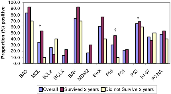
Methods: 23 cases of uterine MMMT identified from the Saskatchewan Cancer Agency (1970-1999) were evaluated. Immunohistochemical expression of Bad, Mcl-1, bcl-x, bak, mdm2, bax, p16, p21, p53, p27, EMA, Bcl-2, Ki67 and PCNA was correlated with clinico-pathological data including survival outcomes.

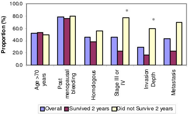
Results: Histopathological examination confirmed malignant epithelial component with homologous (12 cases) and heterologous (11 cases) sarcomatous elements. P53 was strongly expressed (70-95%) in 15 cases and negative in 5 cases. The average survival in the p53+ve cases was 3.56 years as opposed to 8.94 years in p53-ve cases. Overexpression of p16 and Mcl-1 were observed in patients with longer survival outcomes (>2 years). P16 and p21 were overexpressed in the carcinomatous and sarcomatous elements respectively. Cyclin-D1 was focally expressed only in the carcinomatous elements.

Conclusions: Our study supports that a) cell cycle and apoptotic regulatory protein dysregulation is an important pathway for tumorigenesis and b) p53 is an important immunoprognostic marker in MMMT of the uterus.


Figures

Figure 1
Figure 1
Histopathological Evaluation with Immunohistochemical Staining. A: Hematoxylin-eosin stain (original magnification ×250). The star (*) indicates the malignant heterologous component of uterine MMMT. B: Hematoxylin-eosin stain (original magnification ×250). The arrows indicate the malignant epithelial component of uterine MMMT. C: Staining with Bax antibody (original magnification ×250). The expression of Bax antibody is diffuse with the thin arrowhead indicating weak staining, the thick arrowhead indicating medium staining, and the tailed arrow indicating strong staining. D: Staining with p53 antibody(original magnification ×250). The star (*) indicates the negatively stained heterologous sarcomatous element, and the arrow indicates positive staining in the epithelial component. E: Staining with p16 antibody (original magnification ×250). The star (*) indicates the negatively stained region, and the arrow indicates the strong positive staining in the malignant epithelial glands. F: Staining with Cyclin D1 antibody (original magnification ×250). The star (*) indicates the negatively stained heterologous sarcomatous element, while the arrow indicates a focus positive staining in the adjacent epithelial component.
Figure 2
Figure 2
Demographic and Clinical Data in Relation to 2 Year Survival Data. X-axis displays: age, postmenopausal bleeding, homologous elements, stage III/IV, depth of invasion, and metastasis. Y-axis displays: survival outcome data-including overall survival (purple bars), two-year survival (maroon bars), and less than two-year survival (yellow bars). * p < 0.05 based on Fisher's Exact Test.
Figure 3
Figure 3
Cell Cycle and Apoptotic Regulatory Proteins in Relation to 2 Year Survival Data. Statistical significance: *p < 0.05, †p < 0.10.

Similar articles

Cited by

References

    1. Nguyen CP, Levi AW, Montz FJ, Bristow RE. Coexistent Choriocarcinoma and Malignant Mixed Mesodermal Tumor of the Uterus. Gynecol Oncol. 2000;79(3):499–503. doi: 10.1006/gyno.2000.5976. - DOI - PubMed
    1. Robinson-Bennett B, Belch RZ, Han AC. Loss of p16 in Recurrent Malignant Mixed Mullerian Tumors of the Uterus. International Journal of Gynecological Cancer. 2006;16(3):1354–7. doi: 10.1111/j.1525-1438.2006.00411.x. - DOI - PubMed
    1. De Brito PA, Silverberg SG, Orenstein JM. Carcinosarcoma (malignant mixed mullerian (mesodermal) tumor) of the female genital tract: immunohistochemical and ultrastructural analysis of 28 cases. Hum Pathol. 1993;24(2):132–142. doi: 10.1016/0046-8177(93)90291-N. - DOI - PubMed
    1. Silverberg SG, Major FJ, Blessing JA, Fetter B, Askin FB, Liao SY, Miller A. Carcinosarcoma (malignant mixed mesodermal tumor) of the uterus. A Gynecologic Oncology group pathologic study of 203 cases. Int Gynaecol Pathol. 1990;9(1):1–19. doi: 10.1097/00004347-199001000-00001. - DOI - PubMed
    1. Iwasa Y, Haga H, Konishi I, Kobashi Y, Higuchi K, Katsuyama E, Minamiguchi S, Yamabe H. Prognostic factors in uterine carcinosarcoma: a clinicopathologic study of 25 patients. Cancer. 1998;82(3):512–9. doi: 10.1002/(SICI)1097-0142(19980201)82:3<512::AID-CNCR13>3.0.CO;2-4. - DOI - PubMed

Publication types

Substances