. 2010 Jul 21;99(2):553-60.
doi: 10.1016/j.bpj.2010.04.060.

Protein determinants of SNARE-mediated lipid mixing

Affiliations

Protein determinants of SNARE-mediated lipid mixing

Hong Ji et al. Biophys J. .

Abstract

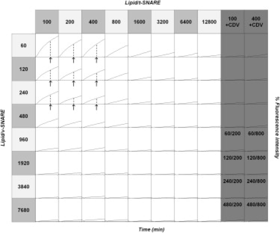
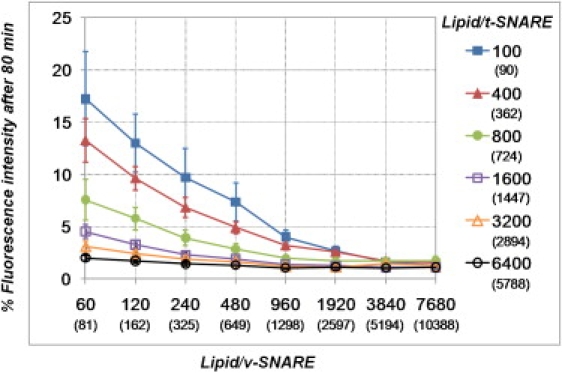
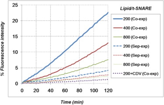
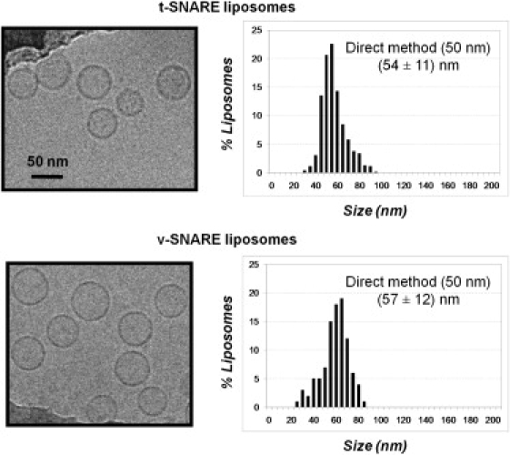
Soluble N-ethylmaleimide sensitive factor attachment protein receptor (SNARE)-mediated lipid mixing can be efficiently recapitulated in vitro by the incorporation of purified vesicle membrane (-v) SNARE and target membrane (t-) SNARE proteins into separate liposome populations. Despite the strong correlation between the observed activities in this system and the known SNARE physiology, some recent works have suggested that SNARE-mediated lipid mixing may be limited to circumstances where membrane defects arise from artifactual reconstitution conditions (such as nonphysiological high-protein concentrations or unrealistically small liposome populations). Here, we show that the previously published strategies used to reconstitute SNAREs into liposomes do not significantly affect either the physical parameters of the proteoliposomes or the ability of SNAREs to drive lipid mixing in vitro. The surface density of SNARE proteins turns out to be the most critical parameter, which controls both the rate and the extent of SNARE-mediated liposome fusion. In addition, the specific activity of the t-SNARE complex is significantly influenced by expression and reconstitution protocols, such that we only observe optimal lipid mixing when the t-SNARE proteins are coexpressed before purification.


Figures

Figure 1
Figure 1
Effect of SNARE density on the kinetics and extent of lipid mixing. Proteoliposomes were prepared by the direct incorporation of SNARE proteins into preformed 50-nm protein-free liposomes. The lipid/protein ratios are theoretical values, derived from starting amounts of protein and lipid. Lipid mixing is measured by monitoring the dequenching of DPPE-NBD lipid probes upon fusion of the fluorescently labeled v-SNARE liposomes with the unlabeled t-SNARE liposomes (x axis range, 0–120 min; y axis range, 0–20% of maximum fluorescence signal; arrows and dashed lines indicate the extent of lipid mixing at t = 80 min, as used in Fig. 2). The last two columns are control experiments in which fusion is inhibited by addition of the cytoplasmic domain of VAMP2 (CDV), which prevents SNAREpin formation by binding to t-SNAREs. The numbers in the last four rows of the control experiments give the v-SNARE and t-SNARE densities used in these reactions. Lipid mixing remains efficient over a wide range of lipid/protein ratios (lipid/t-SNARE ≤ 1600 and lipid/v-SNARE ≤ 480) and, importantly, for SNARE densities consistent with physiology (see main text).
Figure 2
Figure 2
Fusogenicity of SNARE liposomes prepared by the direct incorporation of proteins into preformed 50-nm purely lipidic liposomes (n = 12; error bars indicate standard errors). The extent of lipid mixing is measured as the normalized fluorescence intensity 80 min after initiation of the reaction (Fig. 1, arrows and dashed lines) and plotted against the lipid/v-SNARE ratio (see Fig. S1 for fusion data plotted against the lipid/t-SNARE ratio, and Fig. S3 and Fig. S4 for fusogenicity of SNARE liposomes prepared according to other methods). The lipid/SNARE ratios are depicted either as theoretical (i.e., those expected based on protein and lipid inputs, as in Fig. 1) or as actual (i.e., taking into account average measures of lipid and protein recoveries, as well as protein orientation) values (see section entitled Proteoliposome characterization).
Figure 3
Figure 3
Coexpressed t-SNAREs are more fusogenic than separately expressed t-SNAREs. v-SNARE liposomes 50 nm in size (prepared by the direct method, with lipid/protein = 120) were fused with 50-nm liposomes (direct method) containing t-SNAREs at various surface densities (lipid/protein = 200, 400, or 800) and prepared by two different means: coexpression of Syn1A and SNAP25 as in Figs. 1 and 2 (solid lines) or incubation of separately purified Syn1A and SNAP25 (dashed lines). The extent of lipid mixing is reduced at least fourfold when using separately expressed t-SNARE subunits.
Figure 4
Figure 4
Orientation of SNAREs in the liposome membrane. In this example, proteoliposomes were prepared by direct incorporation of t-SNAREs into 50-nm liposomes. Upon addition of chymotrypsin, t-SNAREs facing outside were proteolyzed, whereas those facing the lumen of the liposomes were protected. Four t-SNARE liposomes with different lipid/protein ratios were exposed to chymotrypsin for 30 min at room temperature and then loaded onto an SDS-PAGE gel, juxtaposed with the same amount of corresponding untreated samples. The percentage of unprotected t-SNAREs, i.e., those exposing their cytosolic domain to the outside, was calculated by comparing the band intensity of the chymotrypsin-treated sample to that of the untreated sample. In this case, ∼75% of the t-SNAREs have their cytoplasmic domain oriented toward the outside of the liposomes. Statistics and results for other SNARE liposomes are displayed in Table 1, Table S1, and Table S2.
Figure 5
Figure 5
Size distribution of SNARE liposomes measured by cryoelectron microscopy. Proteoliposomes were made by direct incorporation of SNAREs into 50-nm protein-free liposomes (as in Figs. 1 and 2). Histograms of t-SNARE and v-SNARE liposomes (lipid/protein = 400 and 60, respectively) were obtained from n = 446 and n = 627 liposomes, respectively; error bars indicate standard deviations. The size distributions of SNARE liposomes prepared by other methods are displayed in Fig. S7.

Similar articles

Cited by

References

    1. Söllner T., Whiteheart S.W., Rothman J.E. SNAP receptors implicated in vesicle targeting and fusion. Nature. 1993;362:318–324. - PubMed
    1. Brunger A.T. Structure and function of SNARE and SNARE-interacting proteins. Q. Rev. Biophys. 2005;38:1–47. - PubMed
    1. Jackson M.B., Chapman E.R. Fusion pores and fusion machines in Ca2+-triggered exocytosis. Annu. Rev. Biophys. Biomol. Struct. 2006;35:135–160. - PubMed
    1. Jahn R., Scheller R.H. SNAREs—engines for membrane fusion. Nat. Rev. Mol. Cell Biol. 2006;7:631–643. - PubMed
    1. Hanson P.I., Roth R., Heuser J.E. Structure and conformational changes in NSF and its membrane receptor complexes visualized by quick-freeze/deep-etch electron microscopy. Cell. 1997;90:523–535. - PubMed

Publication types