

doi: 10.1016/j.bpj.2010.04.040.
Electric field-driven disruption of a native beta-sheet protein conformation and generation of a helix-structure
Affiliations
- PMID: 20643079
- PMCID: PMC2905109
- DOI: 10.1016/j.bpj.2010.04.040
Item in Clipboard
Electric field-driven disruption of a native beta-sheet protein conformation and generation of a helix-structure
Biophys J.
.
Abstract
We demonstrate that an external constant electric field is able to modify the secondary structure of a protein and induce a transition from a beta-sheet into a helix-like conformation. This dramatic change is driven by a global rearrangement of the dipole moments at the amide planes. We also predict electric-field-induced modifications of the intermediate states of the protein.
Copyright (c) 2010 Biophysical Society. Published by Elsevier Inc. All rights reserved.
Figures
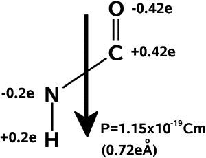
The dipoles of NH and OC in the amide plane give rise to a total dipole moment for each amino acid, which has the value 1.1 × 10⁻²⁹ Cm.
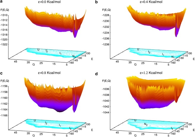
(Color online) Free energy surface of the V3-loop as a function of the configurational energy E and the end-to-end distance Q for different strengths of the external electric field εE0: ε = 0.0, 0.4, 0.8, and 1.2. Local minima labeled as I1 and I2 correspond to intermediates. N1 refers to the native state in absence of field, which becomes metastable (I3) for ε = 0.8. Note the formation of a new global minimum N2 for the field strength ε = 1.2. U corresponds to the unfolded states. The temperature in all cases is T = Tf = 321 K.
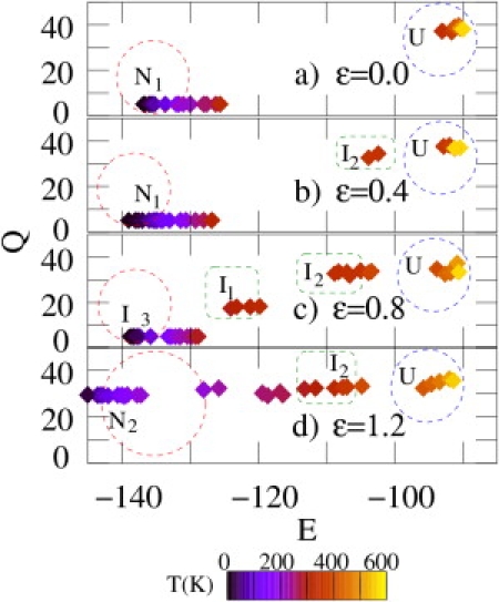
(Color online) Coordinates in the (E, Q)-plane of the conformations yielding the maximal contribution to the partition function for ε = 0.0, 0.4, 0.8, and 1.2 and at different temperatures. The field strength is given by εE0 (see text). Note that for ε = 0.0 the observable structures lie around the point (E = − 135, Q = 5) (β-sheet) while for ε = 1.2 they are located near the point (E = –150, Q = 30) (helix). Dark (blue) and light (yellow) diamonds refer to low and high temperatures, respectively (see temperature scale).
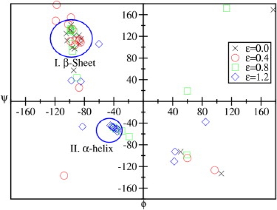
(Color online) Ramachandran plot of the V3-loop for different strengths of the external electric field represented by the dimensionless factor ε (see text) at T = Tf = 321 K. The regions corresponding to helices and β-sheets are indicated.
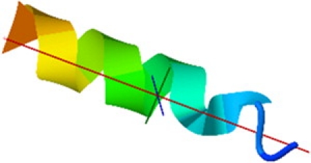
The α-helix structure of the native state for a field strength of εE0 with εE0 = 5.16 × 10⁸.
Similar articles
-
CD4 binding partially locks the bridging sheet in gp120 but leaves the beta2/3 strands flexible.J Mol Biol. 2005 Jul 15;350(3):514-27. doi: 10.1016/j.jmb.2005.05.009. J Mol Biol. 2005. PMID: 15946678
-
Local conformational stability of HIV-1 gp120 in unliganded and CD4-bound states as defined by amide hydrogen/deuterium exchange.J Virol. 2010 Oct;84(19):10311-21. doi: 10.1128/JVI.00688-10. Epub 2010 Jul 21. J Virol. 2010. PMID: 20660185 Free PMC article.
-
Secondary structure of gp160 and gp120 envelope glycoproteins of human immunodeficiency virus type 1: a Fourier transform infrared spectroscopic study.J Virol. 1993 Jun;67(6):3552-60. doi: 10.1128/JVI.67.6.3552-3560.1993. J Virol. 1993. PMID: 8497064 Free PMC article.
-
Pressure-induced transformation of alpha-helix to beta-sheet in the secondary structures of amyloid beta (1-40) peptide exacerbated by temperature.J Biomol Struct Dyn. 2002 Feb;19(4):619-25. doi: 10.1080/07391102.2002.10506768. J Biomol Struct Dyn. 2002. PMID: 11843623
-
HIV gp120: double lock strategy foils host defences.Structure. 1998 Aug 15;6(8):945-9. doi: 10.1016/s0969-2126(98)00096-3. Structure. 1998. PMID: 9739096 Review.
Cited by
-
Dramatic Differences between the Structural Susceptibility of the S1 Pre- and S2 Postfusion States of the SARS-CoV-2 Spike Protein to External Electric Fields Revealed by Molecular Dynamics Simulations.Viruses. 2023 Dec 11;15(12):2405. doi: 10.3390/v15122405. Viruses. 2023. PMID: 38140646 Free PMC article.
-
Electro-opening of a microtubule lattice in silico.Comput Struct Biotechnol J. 2021 Mar 4;19:1488-1496. doi: 10.1016/j.csbj.2021.02.007. eCollection 2021. Comput Struct Biotechnol J. 2021. PMID: 33815687 Free PMC article.
-
Molecular dynamics simulation of the nanosecond pulsed electric field effect on kinesin nanomotor.Sci Rep. 2019 Dec 23;9(1):19721. doi: 10.1038/s41598-019-56052-3. Sci Rep. 2019. PMID: 31873109 Free PMC article.
-
Analysing calcium signalling of cells under high shear flows using discontinuous dielectrophoresis.Sci Rep. 2015 Jul 23;5:11973. doi: 10.1038/srep11973. Sci Rep. 2015. PMID: 26202725 Free PMC article.
-
Nanosecond pulsed electric signals can affect electrostatic environment of proteins below the threshold of conformational effects: The case study of SOD1 with a molecular simulation study.PLoS One. 2019 Aug 27;14(8):e0221685. doi: 10.1371/journal.pone.0221685. eCollection 2019. PLoS One. 2019. PMID: 31454403 Free PMC article.
References
-
- Etienne M.A., Aucoin J.P., Hammer R.P. Stoichiometric inhibition of amyloid β-protein aggregation with peptides containing alternating α,α-disubstituted amino acids. J. Am. Chem. Soc. 2006;128:3522–3523. - PubMed
-
- Kelly J.W. The alternative conformations of amyloidogenic proteins and their multi-step assembly pathways. Curr. Opin. Struct. Biol. 1998;8:101–106. - PubMed
-
- Lynn D.G., Meredith S.C. Review: Model peptides and the physicochemical approach to β-amyloids. J. Struct. Biol. 2000;130:153–173. - PubMed
-
- Wada A. The α-helix as an electric macro-dipole. Adv. Biophys. 1976;9:1–63. - PubMed
MeSH terms
Substances
LinkOut - more resources
Full Text Sources
