. 2010 Jul 21;99(2):685-94.
doi: 10.1016/j.bpj.2010.04.058.

Backbone trace of partitivirus capsid protein from electron cryomicroscopy and homology modeling

Affiliations

Backbone trace of partitivirus capsid protein from electron cryomicroscopy and homology modeling

Jinghua Tang et al. Biophys J. .

Abstract

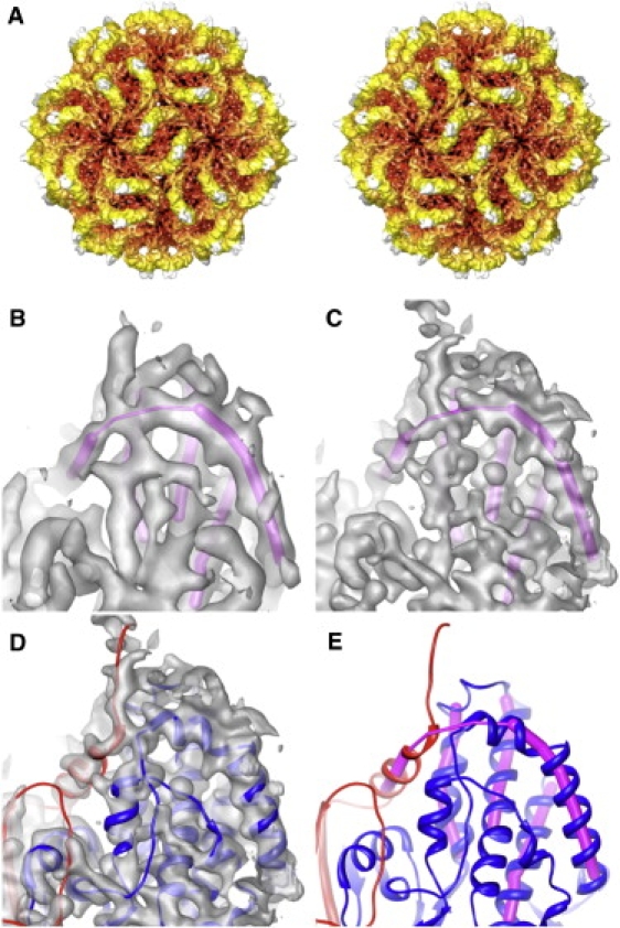
Most dsRNA viruses have a genome-enclosing capsid that comprises 120 copies of a single coat protein (CP). These 120 CP subunits are arranged as asymmetrical dimers that surround the icosahedral fivefold axes, forming pentamers of dimers that are thought to be assembly intermediates. This scheme is violated, however, in recent structures of two dsRNA viruses, a fungal virus from family Partitiviridae and a rabbit virus from family Picobirnaviridae, both of which have 120 CP subunits organized as dimers of quasisymmetrical dimers. In this study, we report the CP backbone trace of a second fungal partitivirus, determined in this case by electron cryomicroscopy and homology modeling. This virus also exhibits quasisymmetrical CP dimers that are connected by prominent surface arches and stabilized by domain swapping between the two CP subunits. The CP fold is dominated by alpha-helices, although beta-strands mediate several important contacts. A dimer-of-dimers assembly intermediate is again implicated. The disordered N-terminal tail of each CP subunit protrudes into the particle interior and likely interacts with the genome during packaging and/or transcription. These results broaden our understanding of conserved and variable aspects of partitivirus structure and reflect the growing use of electron cryomicroscopy for atomic modeling of protein folds.


Figures

Figure 1
Figure 1
PsV-F capsid structure. (A) Stereo view along a 2f direction of a cryoTEM reconstruction at 6.0 Å resolution. The particle is radially depth-cued, from red (low radii) to white (high radii). (B and C) The same magnified view of cryoTEM reconstructions respectively calculated at 6.0 and 4.7 Å resolution. Superimposed in magenta is an interpretation of the main-chain directionality solely from these maps. (D) Crystal-derived atomic model of PsV-F CP dimer (8) fitted into the 4.7 Å map. (E) Atomic model of PsV-F CP dimer with the two chains shown in red and blue, indicating the close contact between these two chains that leads to the wrong interpretation of main-chain directionality in B and C.
Figure 2
Figure 2
Resolution estimation of the PsV-F cryoTEM map based on crystallographic maps. (A) CC plots comparing the cryoTEM map (EM) with the 3.3 Å x-ray crystallographic map (xtal) calculated at different resolution (res) limits, in Å. (B) CC plots comparing the final cryoTEM map (EM) with different versions of the PsV-F atomic model.
Figure 3
Figure 3
PsV-S capsid structure. (A) Stereo view along a 2f direction of a cryoTEM reconstruction at ∼4.5 Å resolution (estimated). The particle is radially depth-cued, from green (low radii) to red (high radii). (B) The CPA-CPB dimer assumes different directionality (green lines; angle measurements estimated at bottom) in PsV-S (left) and -F (right). Both particles are viewed along a 2f direction. Symmetry elements are evident from the icosahedral line drawing superimposed on each particle. The approximate boundaries of the putative assembly intermediate, a dimer of CPA-CPB dimers, are outlined by a red diamond in each virus. (C) Magnified views of two regions of the PsV-S cryoTEM map, represented as a wire mesh and with positions of Cα atoms modeled as cyan spheres and connected by red lines to indicate chain path. For C, the map is displayed at a high density-contour level (2.0 σ) to exclude low-density features.
Figure 4
Figure 4
Secondary-structure assignments. The secondary structures of PsV-S are shown above the sequence (shell domain in red, arch domain in green), and those of PsV-F below the sequence (shell domain in magenta, arch domain in bluish-green). The predicted secondary structure of PsV-S is shown in gray at top. α-helices are represented by rods, β-strands by arrows, random coils by solid lines, and disordered regions by broken lines.
Figure 5
Figure 5
3D structures of PsV-S and -F CP dimers. (A) Side view and (B) inside-out view of a CPA-CPB dimer of (left) PsV-S and (right) -F. The color scheme is identical to that in Fig. 4. The two subunits from a CP dimer are colored in different shades for better differentiation. Secondary structures are labeled as defined in Fig. 4. (C) Topology diagram of (left) PsV-S and (right) -F. Secondary structures are labeled by numbers only.
Figure 6
Figure 6
Genome organization and transcription. The PsV-S cryoreconstruction was computed to a resolution cutoff of (A) 10 Å or (B and C) 8 Å to highlight less-ordered components in the particle interior. Density-contour levels for the three panels were 1.05, 0.85, and 1.5 σ. The cryoTEM map is represented by a wire mesh, within which the Cα backbone of the PsV-S CP subunits (CPA, red; CPB, yellow) are fitted. (A) Internal genomic RNA densities. Cyan curves highlight the two RNA density layers. (B) Rod-like densities on the inner capsid surface. The disordered N-termini of CPA and CPB are highlighted by red and yellow stars, respectively. (C) PsV-S capsid structure around the 5f symmetry axis. The N-terminus of one CPB subunit is highlighted by a yellow circle.

Similar articles

Cited by

References

    1. Mertens P. The dsRNA viruses. Virus Res. 2004;104:3–13. - PubMed
    1. Grimes J.M., Burroughs J.N., Stuart D.I. The atomic structure of the bluetongue virus core. Nature. 1998;395:470–478. - PubMed
    1. Naitow H., Tang J., Johnson J.E. L-A virus at 3.4 Å resolution reveals particle architecture and mRNA decapping mechanism. Nat. Struct. Biol. 2002;9:725–728. - PubMed
    1. Castón J.R., Ghabrial S.A., Carrascosa J.L. Three-dimensional structure of Penicillium chrysogenum virus: a double-stranded RNA virus with a genuine T=1 capsid. J. Mol. Biol. 2003;331:417–431. - PubMed
    1. Coulibaly F., Chevalier C., Rey F.A. The birnavirus crystal structure reveals structural relationships among icosahedral viruses. Cell. 2005;120:761–772. - PubMed

Publication types

Substances

LinkOut - more resources