. 2010 Oct 1;346(1):39-53.
doi: 10.1016/j.ydbio.2010.07.011. Epub 2010 Jul 17.

The ATP-sensitive K(+)-channel (K(ATP)) controls early left-right patterning in Xenopus and chick embryos

Affiliations

The ATP-sensitive K(+)-channel (K(ATP)) controls early left-right patterning in Xenopus and chick embryos

Sherry Aw et al. Dev Biol. .

Abstract

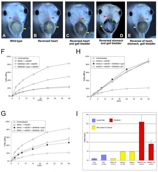
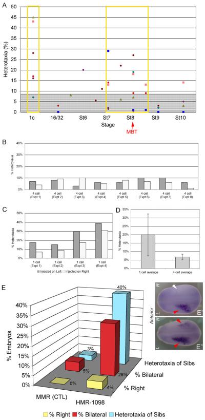
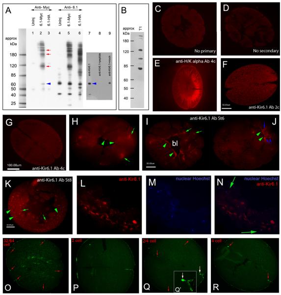
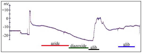
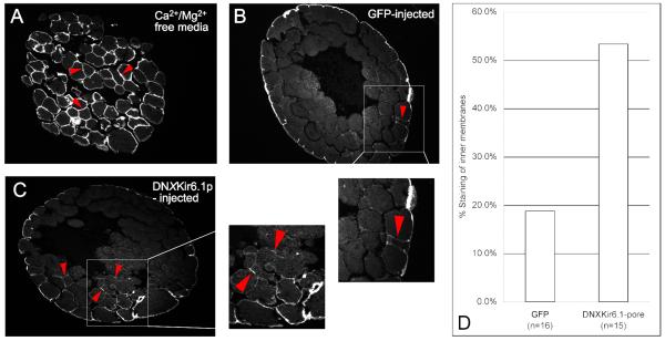
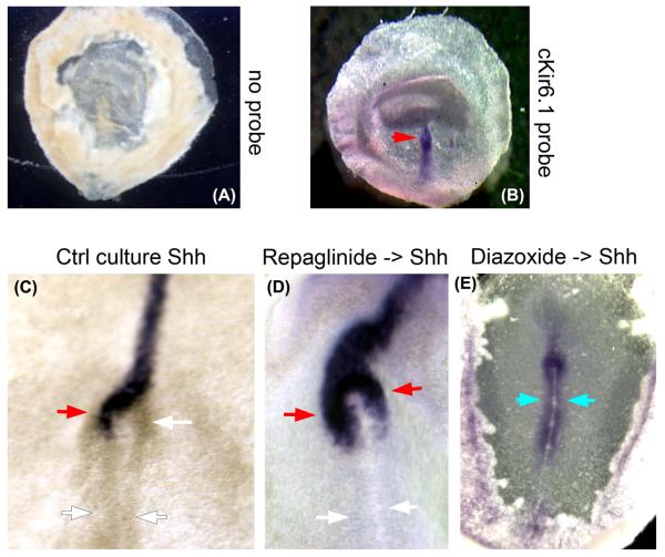
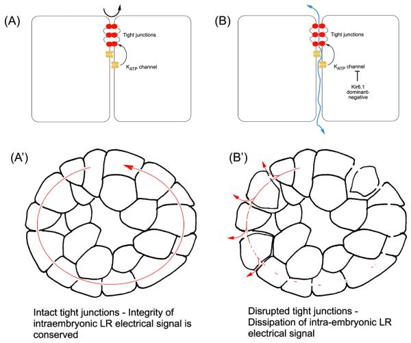
Consistent left-right asymmetry requires specific ion currents. We characterize a novel laterality determinant in Xenopus laevis: the ATP-sensitive K(+)-channel (K(ATP)). Expression of specific dominant-negative mutants of the Xenopus Kir6.1 pore subunit of the K(ATP) channel induced randomization of asymmetric organ positioning. Spatio-temporally controlled loss-of-function experiments revealed that the K(ATP) channel functions asymmetrically in LR patterning during very early cleavage stages, and also symmetrically during the early blastula stages, a period when heretofore largely unknown events transmit LR patterning cues. Blocking K(ATP) channel activity randomizes the expression of the left-sided transcription of Nodal. Immunofluorescence analysis revealed that XKir6.1 is localized to basal membranes on the blastocoel roof and cell-cell junctions. A tight junction integrity assay showed that K(ATP) channels are required for proper tight junction function in early Xenopus embryos. We also present evidence that this function may be conserved to the chick, as inhibition of K(ATP) in the primitive streak of chick embryos randomizes the expression of the left-sided gene Sonic hedgehog. We propose a model by which K(ATP) channels control LR patterning via regulation of tight junctions.


Figures

Figure 1
Figure 1. The KATP channel is necessary for correct left-right patterning
A) Untreated embryo exhibiting normal situs (situs solitus). B-E) Embryos treated with KATP channel blocker HMR-1098 (1.45mM) (Dhein et al., 2000; Edwards et al., 2009; Jovanovic and Jovanovic, 2005; Light et al., 2001; Suzuki et al., 2003) from Stage 1 cell to Stage 16 (then washed out into 0.1x MMR and allowed to develop to Stage 46 before being scored for organ situs) exhibit heterotaxia, including B) reversed heart C) reversed heart and gall bladder D) reversed stomach and gall bladder E) Reversed heart, stomach and gall bladder. F-G) Rubidium flux assays of COSm6 cells expressing various KATP subunits. F) xKir6.1 with mouse SUR1 (mSUR1) dramatically increased the rate of efflux compared to untransfected cells by about four-fold, suggesting the formation of function KATP channels. Expression of DNxKir6.1-pore or DNxKir6.1-ER with mouse SUR1 (mSUR1) show that the ER mutant is able to conduct K+ currents with mSUR1, whilst the pore mutant is non-conductive. G,H) DNxKir6.1-pore and DNxKir6.1-ER knock down activity of KATP channels consisting of WTxKir6.1 and mouse SUR1 (F), but not those consisting of mouse Kir6.2 and mouse SUR1 (G). I) Injection of dominant-negatives against xKir6.1 causes significant levels of heterotaxia, but not injection of GFP, or dominant-negative mRNAs against other Kir channels DNKir2.1 (Zobel et al., 2003), DNKir2.2 (Zobel et al., 2003) and DNKir2.3 (Bannister et al., 1999).
Figure 2
Figure 2. The KATP channel functions in both early and late cleavage in LR patterning, and signals to the left-sided Nodal signaling cascade
A) Embryos were incubated in KATP channel blocker HMR-1098 starting from the indicated stages, and washed out at ~Stage 19. Each differently-colored dot represents embryos from a different mother. The gray shaded area on the chart indicates 9% heterotaxia, which is the level of heterotaxia required in a dish of 100 embryos to achieve significance of heterotaxia over a dish of controls with 2% heterotaxia in a chi-squared test. Significant rates of heterotaxia were observed when embryos were incubated in HMR-1098 starting from one cell, and from about Stage 7 to Stage 8.5. MBT, mid-blastula transition (see text for details). B) Dominant-negative xKir6.1-pore does not cause significantly different rates of heterotaxia when it is injected on the left versus the right side of the embryo at four cell (difference is not significant, p>0.6 by paired t-test). C) When asymmetric injections were repeated at one cell, it was found that injection on the left gives a consistently higher rate of heterotaxia than injection on the right, and left injected embryos exhibit a consistently higher rate of heterotaxia than right-injected embryos in every experiment (p<0.01 by paired t-test). D) Injections in one half of the embryo at one cell causes approximately 3X as many embryos with heterotaxia as compared to injections in one half of the embryo at four cell, showing that the KATP channel has a role in LR patterning at the earliest cleavage stages. E) KATP channel blocker HMR-1098 randomizes Nodal. Wild-type embryos typically exhibit Nodal expression only on the left side of the embryo (E′). Control embryos incubated in 0.1XMMR media alone exhibited only 4% incorrect Nodal staining (right-sided or bilateral staining), and control sibling embryos exhibited a similar 3% rate of background heterotaxia. However, embryos incubated in HMR-1098 exhibited 33% right-sided (image not shown) and bilateral Nodal staining (E″), compared to the 40% rate of heterotaxia seen in sibling embryos incubated in HMR-1098 (E).
Figure 3
Figure 3. Immunolocalization of Xenopus Kir6.1 and SUR2A
A) Western blotting with anti-myc and anti-Kir6.1 antibodies against lysates from Stage 11 embryos injected at one cell with xKir6.1-myc mRNA. The blot confirms that both antibodies recognize products with molecular weights at multiples of 60kDa (lanes 2 and 5). Although this is slightly larger than the up to ~51kDa size recognized for Kir6.1 in other studies (Foster et al., 2008; Suzuki et al., 1997), it runs at the same size as the Kir6.1-myc construct probed with anti-Myc (blue arrow, lane 2), confirming that the anti-Kir6.1 antibody recognizes xKir6.1 protein. The higher molecular weight bands were not observed in lysates from uninjected embryos (lanes 1 and 4). Lysates of uninjected one-cell Xenopus embryos shows that 1-cell embryos contain maternal xKir6.1 protein (lanes 4 and 7, blue arrow). Pre-incubation of the antibody with Kir6.1 peptide (lane 8), but not with a Kir6.2 peptide (lane 9), abolished immunoreative bands. Anti-myc recognized a faint band at about 45-kDa in uninjected embryos (lane 1); this may be endogenous c-myc. B) A SUR2 antibody recognizes several bands in one cell Xenopus lysates, including a 130 kDa band, approximately the size of SUR2 (150 kDa). C-K) Immunofluorescence with anti-Kir6.1 antibody on sections from early to late cleavage stage in Xenopus embryos. C) Control sections without primary or (D) secondary antibodies exhibit low level staining and low levels of yolk autofluorescence. E) Positive control anti-H/K-ATPase antibody shows expected left-right asymmetric localization. F, G) At two to four cell stage, xKir6.1 exhibits low levels of expression and indistinct localization. H) At Stage 6, xKir6.1 is localized to intracellular domains (green arrows) and to isolated regions on plasma membranes facing the blastocoel (green arrowheads). I, J) At stage 7, Kir6.1 is localized to intracellular domains (I, green arrows) and membranes on the basal face of animal cap cells lining the blastocoel (J, green arrowheads). Intercellular punctate staining is also observed (J, blue arrows). K) At Stage 8, xKir6.1 remains localized to intracellular domains (K, green arrows) and to plasma membrane in many parts of the embryo (K, green arrowheads). L-N) When intracellular domains recognized by anti-Kir6.1 (L) are co-localized with nuclear DNA (M), they appear to lie peripheral to the nucleus (N), suggestive of endoplasmic reticulum localization. O-R) Immunofluorescence with anti-SUR2A antibody in early cleavage Xenopus embryos show punctate, tight junction-like staining at from two cell to stage 32/64 cell. Q′ is a close up of the staining at the junction of a cleaving two to four cell embryo, indicated with a white arrow in Q.
Figure 4
Figure 4. KATP channel activity in the early cleavage Xenopus embryo is detectable in a few surface blastomeres
Whole cell electrophysiological recordings from random locations in developing cleavage stage Xenopus embryos revealed three of 20 blastomeres impaled that responded in a manner typical of KATP channels, exhibiting the predicted response to combinations of the treatments diazoxide, azide and glibenclamide, e.g. hyperpolarizing in response to azide, hyperpolarizing in response to diazoxide, depolarizing in response to glibenclamide and repolarizing upon removal of glibenclamide. Figure 4A shows a representative trace of one of the three responding blastomeres.
Figure 5
Figure 5. Dominant-negative DNxKir6.1-pore against the KATP channel changes tight junction properties of early cleavage stage Xenopus embryos
A tight-junction and membrane impermeable biotin that labels surface proteins allowed probing of tight junction integrity in Xenopus embryos (Merzdorf et al., 1998). A) Incubation of embryos in Ca2+ and Mg2+-free media caused breaking of tight junctions and extensive labeling of inner membranes. B) Control embryos injected with Venus-GFP mRNA showed faint staining of inner membranes, but not more than one cell layer deep (red arrowhead and inset). C) Embryos injected with DNxKir6.1-pore showed extensive staining of inner membranes, often several cell layers deep (red arrowheads and inset). D) In total, 3/16 embryos (19%) injected with control GFP mRNA exhibited staining of membranes one cell layer or more deep, and 8/15 (53%) of embryos injected with DNxKir6.1-pore mRNA exhibited staining of membranes one cell layer or more deep.
Figure 6
Figure 6. KATP function in the LR pathway is conserved in chick embryogenesis
A, B) Chick Kir6.1 is transcribed throughout the early primitive streak in a symmetrical manner, at the time during which differential membrane voltage gradients are establishing asymmetric gene expression (Levin et al., 2002). C) Control embryos exposed to vehicle alone exhibited 94% left-sided Shh. D) 29% of embryos exposed bilaterally to 10.6 μM KATP blocker repaglinide (Dabrowski et al., 2001; Gromada et al., 1995) exhibited randomized expression of Shh (p<0.01 by chi-squared test). E) Bilateral exposure to 200 μM KATP activator diazoxide (D'Hahan et al., 1999) randomized Shh, also inducing a unique phenotype not known to be observed after any other treatment: expansion of Shh expression posteriorly from the node into the mid-streak.
Figure 7
Figure 7. A model of how disruption of tight junctions by the KATP channel might cause defects in LR patterning
A) In a normal embryo, tight junctional integrity is conserved in the presence of functional KATP channels, preventing paracellular flow and leakage (black arrow). A′) In the context of the embryo, functionally robust tight junctional seals would support proper gap junction function and prevent short circuit caused by paracellular leakage, allowing conservation of the intraembryonic LR electrical signal (see text for details). B) The disruption of KATP channels by xKir6.1 dominant-negatives causes loss of tight junctional integrity and hence paracellular leakage (blue arrow), which would alter both transepithelial potentials, and secondarily, transmembrane voltage gradients in the blastomeres. B′) Disruption of tight junctions is predicted to cause rapid dissipation of the intraembryonic LR electrical signal.

Similar articles

Cited by

References

    1. Adams DS, Levin M. Inverse drug screens: a rapid and inexpensive method for implicating molecular targets. Genesis. 2006a;44:530–40. - PMC - PubMed
    1. Adams DS, Levin M. Strategies and techniques for investigation of biophysical signals in patterning. In: Whitman M, Sater AK, editors. Analysis of Growth Factor Signaling in Embryos. Taylor and Francis Books; 2006b. pp. 177–262.
    1. Adams DS, Robinson KR, Fukumoto T, Yuan S, Albertson RC, Yelick P, Kuo L, McSweeney M, Levin M. Early, H+-V-ATPase-dependent proton flux is necessary for consistent left-right patterning of non-mammalian vertebrates. Development. 2006a;133:1657–71. - PMC - PubMed
    1. Adams DS, Robinson KR, Fukumoto T, Yuan S, Albertson RC, Yelick P, Kuo L, McSweeney M, Levin M. Early, H+-V-ATPase-dependent proton flux is necessary for consistent left-right patterning of non-mammalian vertebrates. Development. 2006b;133:1657–1671. - PMC - PubMed
    1. Albrieux M, Villaz M. Bilateral asymmetry of the inositol trisphosphate-mediated calcium signaling in two-cell ascidian embryos. Biology of the Cell. 2000;92:277–84. - PubMed

Publication types

MeSH terms

LinkOut - more resources