. 2010 Sep 10;402(1):194-209.
doi: 10.1016/j.jmb.2010.07.022. Epub 2010 Jul 17.

Structural evidence that peroxiredoxin catalytic power is based on transition-state stabilization

Affiliations

Structural evidence that peroxiredoxin catalytic power is based on transition-state stabilization

Andrea Hall et al. J Mol Biol. .

Abstract

Peroxiredoxins (Prxs) are important peroxidases associated with both antioxidant protection and redox signaling. They use a conserved Cys residue to reduce peroxide substrates. The Prxs have a remarkably high catalytic efficiency that makes them a dominant player in cell-wide peroxide reduction, but the origins of their high activity have been mysterious. We present here a novel structure of human PrxV at 1.45 A resolution that has a dithiothreitol bound in the active site with its diol moiety mimicking the two oxygens of a peroxide substrate. This suggests diols and similar di-oxygen compounds as a novel class of competitive inhibitors for the Prxs. Common features of this and other structures containing peroxide, peroxide-mimicking ligands, or peroxide-mimicking water molecules reveal hydrogen bonding and steric factors that promote its high reactivity by creating an oxygen track along which the peroxide oxygens move as the reaction proceeds. Key insights include how the active-site microenvironment activates both the peroxidatic cysteine side chain and the peroxide substrate and how it is exquisitely well suited to stabilize the transition state of the in-line S(N)2 substitution reaction that is peroxidation.


Figures

Figure 1
Figure 1
Electron density for bound DTT in the active site. Shown is the electron density in the active site (A) before including DTT in the model and (B) after the final round of refinement of the model containing DTT (HsPrxV·DTT). A peptide segment including the CP and the two residues prior to it is shown in sticks and colored by atom (oxygen = red, nitrogen = blue, sulfur = yellow), and electron density is shown as mesh: 2Fo-Fc density is contoured at 1.5 ρrms (pale blue) and 6.5 ρrms (dark blue), and Fo-Fc density is contoured at 4 ρrms (green). Panel B inset: An overlay of the final HsPrx·DTT structure (dark grey carbons) with the published HsPrx·BEZ structure (light grey carbons; PDB entry 1HD2) in the same orientation as the main panel.
Figure 2
Figure 2
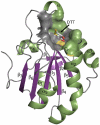
The HsPrx·DTT structure. The protein backbone ribbon is shown with α-helices (green), β-strands (purple), coils/turns (grey), and the CP residue (yellow) highlighted. The bound DTT molecule (colored by atom, oxygen = red, sulfur = yellow) is shown as sticks, and the surface of the active site pocket into which it binds is shown. Secondary structures are labeled according to the common fold of all Prxs; the short helix between α4 and α5 is not universally conserved in the Prx fold and is labeled simply as “α”.
Figure 3
Figure 3
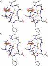
Stereoview of the HsPrx·DTT active site with bound DTT. Shown are the CP-loop and the first turn of α2 plus the conserved Arg colored by atom as in Figure 1. Hydrogen bonding (cyan dotted lines) important for DTT binding and the SP-OA interaction (orange dashed line) are indicated. The four residues conserved in all Prxs and the His that interacts with the active site Arg are labeled.
Figure 4
Figure 4
Two distinct positions for the conserved Arg in the Prx active site. (A) The overlaid active sites from all ligand-bound Prx structures (Table 1) are shown with side chains (shown as sticks) for the CP, the conserved active site Arg, the supporting Glu/Gln/His residue and for those that contain it, a second Arg (indicated by an asterisk) of the Arg-Glu-Arg hydrogen bonding network. The carbon atoms are colored according to the position of the Arg, light grey for position I and purple for position II; oxygen (red), nitrogen (blue) and sulfur (yellow) are colored by atom. The distinct positioning of the conserved active site Arg correlates with the presence of the second Arg residue. (B) Same as panel A but including all FF Prx structures. The overlay includes 179 chains from 32 pdb entries (3A2V, 3A2W, 1QMV, 2Z9S, 2PN8, 1UUL, 2I81, 2V2G, 2V32, 2V41, 1XCC, 2CV4, 1X0R, 3A5W, 1HD2, 1H4O, 1OC3, 1URM, 2VL2, 1TP9, 2VL3, 1NM3, 1XIY, 3HVV, 1Y25, 1PSQ, 2YZH, 2CX4, 1XXU, 3MNG, 3DRN, 3GKM).
Figure 5
Figure 5
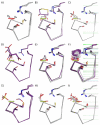
Overlays of ligands that bind in the Prx active site. In all panels, a peptide segment containing the CP-loop is shown as a ribbon in the same orientation as in Figure 3. Side chains are shown for the conserved Pro, Thr, CP and Arg, and ligands are shown as sticks with coloring as in Figure 4. (A) H2O2-bound structure with OA, OB and SP atoms identified. In the remaining panels, a bound H2O2 molecule (transparent pale green) is shown for reference. Panels B through I feature the following ligands: (B) DTT and ethanediol; (C) glycerol; (D) citrate; (E) acetate; (F) benzoate; (G) sulfate and formate; (H) ethanediol binding as only OA; (I) glycerol binding as only OA. The structures included in each panel are listed in Table 1.
Figure 6
Figure 6
Summary of peroxide-mimics binding in the Prx active site. (A) The oxygen atoms (red spheres) mimicking OA and OB from all ligands shown in Figure 5. (B) Active site water molecules (red spheres) from unliganded FF structures. In both panels, the CP-loop and conserved Arg are shown as sticks in the same orientation as Figure 3 and colored by atom type. As in Figure 5, a reference H2O2 molecule (pale green) is included for reference. In this figure the CP-loop is extended by one residue to emphasize it is the first turn of helix α2 by showing the first hydrogen bond of the helix (cyan). Waters shown are from pdb codes 1X0R, 1QMV, 2PN8, 1XXU, 1PSQ, 1XIY, 3A5W, 1CXX and 1N8J.
Figure 7
Figure 7
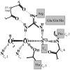
Interactions in the Prx active site Michaelis complex. Shown is a schematic drawing of the peroxide-bound active site emphasizing the hydrogen bonding interactions present. The in-line geometry for SP to attack OA for the peroxidatic SN2 reaction (bold dashed line) and key hydrogen bonding interactions (pale dashed, dotted, and solid lines) are indicated. The view is as in Figures 3, 5, 6 and 8. Backbone atoms in the CP-loop are identified by their residue position relative to CP. The negative charge on the CP-thiolate is indicated.
Figure 8
Figure 8
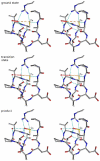
Reaction path for the peroxidatic SN2 reaction. (A) The Michaelis complex structure as seen in ApTpx·H2O2 chain F (PDB entry 3A2V20) shows how the active site aligns the peroxide substrate for an in-line attack from the CP-thiolate (orange dashed line). (B) Simplistically-derived model for the transition state (see text) showing the partial SP-OA (orange dashed line) and OA-OB (red dashed line) bonds. The SP-OA and OA-OB distances are 2.6 and 2.4 Å, respectively. (C) The product complex as seen at ~25% occupancy in the rerefined ApTpx·H2O2 chain F (see text). The SPOAH and OB hydroxide leaving group (red sphere) are shown. Stabilizing active site hydrogen bonds (cyan dotted lines) are shown in all panels.

References

    1. Karplus PA, Hall A. Structural survey of the peroxiredoxins. In: Flohé L, Harris JR, editors. Peroxiredoxin Systems. Springer; New York: 2007. pp. 41–60. - PubMed
    1. Hall A, Karplus PA, Poole LB. Typical 2-Cys peroxiredoxins--structures, mechanisms and functions. Febs J. 2009;276:2469–77. - PMC - PubMed
    1. Chae HZ, Robison K, Poole LB, Church G, Storz G, Rhee SG. Cloning and sequencing of thiol-specific antioxidant from mammalian brain: alkyl hydroperoxide reductase and thiol-specific antioxidant define a large family of antioxidant enzymes. Proc Natl Acad Sci U S A. 1994;91:7017–21. - PMC - PubMed
    1. Flohe L, Budde H, Hofmann B. Peroxiredoxins in antioxidant defense and redox regulation. Biofactors. 2003;19:3–10. - PubMed
    1. Akerman SE, Muller S. Peroxiredoxin-linked detoxification of hydroperoxides in Toxoplasma gondii. J Biol Chem. 2005;280:564–70. - PubMed

Publication types

LinkOut - more resources