. 2010 Jul 20;19(1):138-47.
doi: 10.1016/j.devcel.2010.06.008.

Epidermal wound repair is regulated by the planar cell polarity signaling pathway

Affiliations

Epidermal wound repair is regulated by the planar cell polarity signaling pathway

Jacinta Caddy et al. Dev Cell. .

Erratum in

  • Dev Cell. 2010 Aug 17;19(2):353. Parekh, Vishwas [added]

Abstract

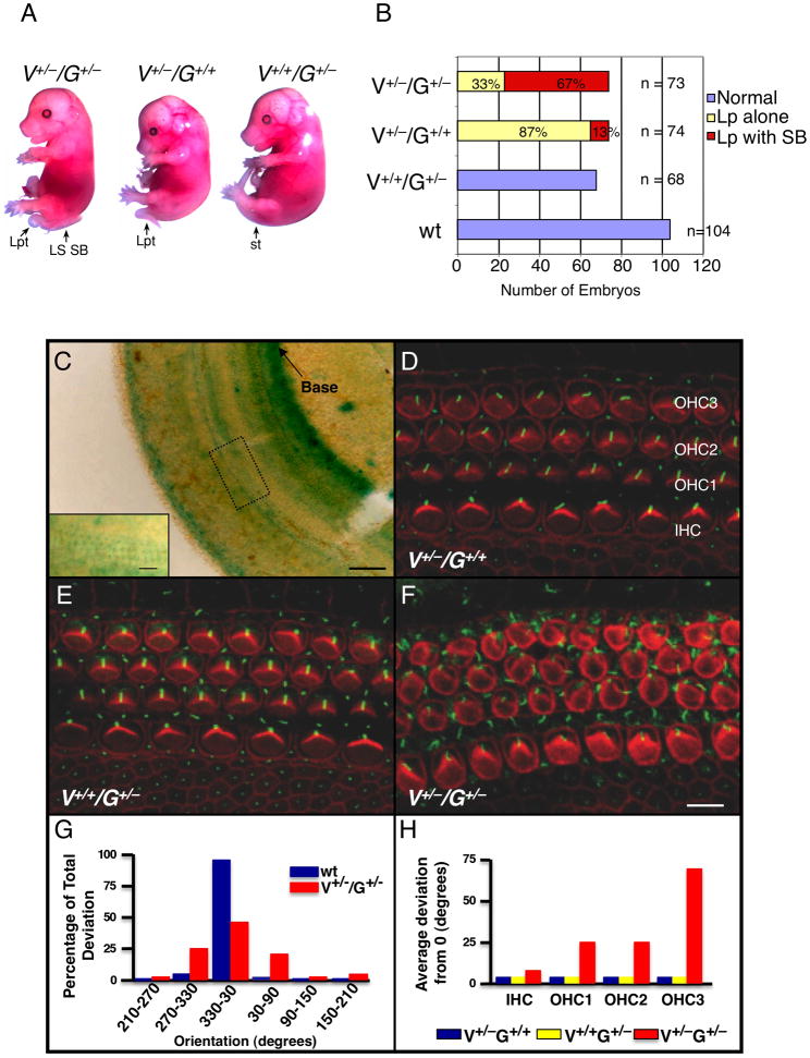
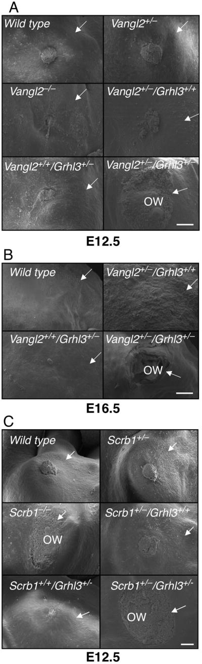
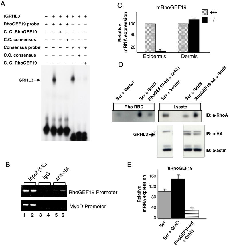
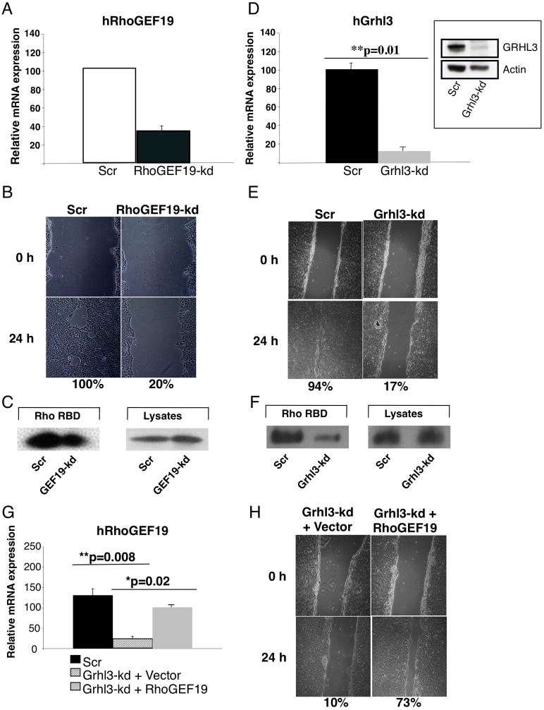
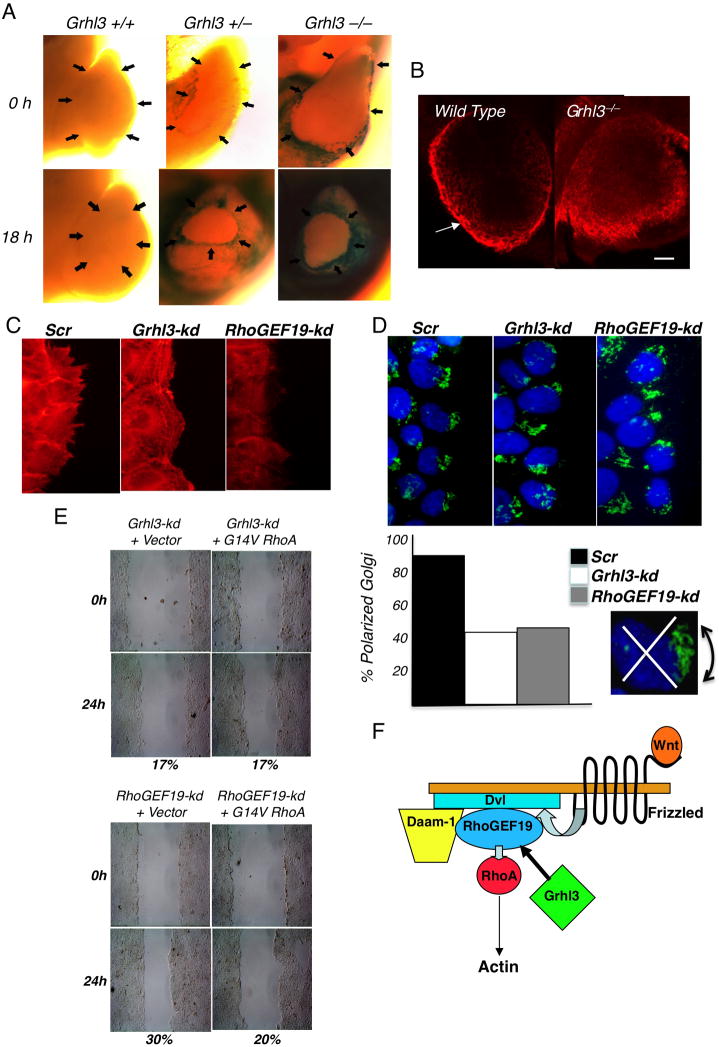
The mammalian PCP pathway regulates diverse developmental processes requiring coordinated cellular movement, including neural tube closure and cochlear stereociliary orientation. Here, we show that epidermal wound repair is regulated by PCP signaling. Mice carrying mutant alleles of PCP genes Vangl2, Celsr1, PTK7, and Scrb1, and the transcription factor Grhl3, interact genetically, exhibiting failed wound healing, neural tube defects, and disordered cochlear polarity. Using phylogenetic analysis, ChIP, and gene expression in Grhl3(-)(/-) mice, we identified RhoGEF19, a homolog of a RhoA activator involved in PCP signaling in Xenopus, as a direct target of GRHL3. Knockdown of Grhl3 or RhoGEF19 in keratinocytes induced defects in actin polymerization, cellular polarity, and wound healing, and re-expression of RhoGEF19 rescued these defects in Grhl3-kd cells. These results define a role for Grhl3 in PCP signaling and broadly implicate this pathway in epidermal repair.


Figures

Figure 1
Figure 1
Genetic interaction between Grhl3 (G) and Vangl2 (V) in neural tube closure and cochlear stereociliary bundle orientation. (A) E15.5 littermates from Vangl2+/−/Grhl3+/−matings. Lp: loop tail; LS SB: lumbo-sacral spina bifida; st: straight tail. (B) Summary of the incidence of spina bifida in the Vangl2/Grhl3 compound heterozygotes. A total of 319 embryos from 46 separate litters of Vangl2 and Grhl3 heterozygote matings were analysed. (C) Section of an entire cochlear duct from a Grhl3+/− embryo at E18.5 stained with β-galactosidase. Inset is high magnification of the boxed region, illustrating the LacZ expressing hair cell layers. (D-F) Luminal surface of the organ of Corti in the middle turn of cochleae from E18.5 embryos with the indicated genotypes. Stereociliary bundles (red) and kinocilia (green) on IHC and OHC are uniformly orientated in (D) and (E). Bundles are non-uniform in (F) with some rotated by greater than 900. The kinocilia are also frequently mispositioned in (F). (G) Distribution histograms of OHC3 bundle orientation in wild type and Vangl2+/− /Grhl3+/− cochleae. Wild type OHCs orientations are largely confined to a 600 segment centered on a line parallel to the neural-abneural axis. In contrast the distributions are noticeably broader in Vangl2+/− /Grhl3+/− mice. (H) Average deviations from a line parallel to the neural-abneural axis for all hair cell layers in the genotypes shown in (D-F). See also Figure S1.
Figure 2
Figure 2
Failed wound healing in PCP mutant mice. SEM of a hind limb amputation wound in embryos with the stated genotypes at (A) E12.5; (B) E16.5; and (C) E12.5. All images are representative of at least six embryos of each genotype. Wounds were classified as open if the residual defect after 24 hours culture was >80% of the original wound diameter. Arrows point to the boundary of the wound. ow - open wound. Scale = 100 μm. See also Figure S2 and Table S1.
Figure 3
Figure 3
RhoGEF19 is a direct target gene of GRHL3. (A) EMSA of recombinant (r) GRHL3 binding to the RhoGEF19 promoter. A 100-fold molar excess of unlabelled cold competitor probe was added in the indicated lanes. The migration of the specific GRHL3/DNA complexes is arrowed. (B) ChIP analysis of GRHL3 on the RhoGEF19 promoter. Chromatin from HEK 293T cells transfected with empty vector (lane 5) or HA-tagged Grhl3 (lane 6) was immunoprecipitated using antisera to the HA-tag. As negative controls, we used pre-immune sera (lanes 3 and 4). Lane 1 and 2 shows the input chromatin. The immunoprecipitated chromatin was amplified with RhoGEF19, or control MyoD primers. (C) Q-PCR of RhoGEF19 expression in wild type (hatched boxes) and Grhl3−/− (closed boxes) E18.5 epidermis and dermis. Bars represent standard errors. The HPRT levels served as a reference. (D, E) GRHL3 induced RhoA activation in HEK 293T cells is mediated through increased RhoGEF19 expression. HA-tagged Grhl3 was transfected into HEK 293T cells transduced with an shRNA to RhoGEF19, or a Scrambled control shRNA (Scr), and expression of HA-GRHL3 was confirmed by Western blot (IB:α-HA, α-actin loading control). GTP-Rho was precipitated using GST-RBD and detected by anti-RhoA antibody (IB:αRhoA). Total RhoA was also detected in lysates with anti-RhoA antibody. HA-GRHL3 is arrowed. *non-specific band with HA antibody. Expression of human RhoGEF19 in Scr control cells, Scr cells expressing HA-GRHL3, and RhoGEF19-kd cells expressing HA-GRHL3 was determined by Q-PCR. See also Figure S3.
Figure 4
Figure 4
Re-expression of RhoGEF19 rescues the wound healing defect in Grhl3-deficient keratinocytes. (A) Q-PCR of RhoGEF19 expression in Scr and RhoGEF19-kd HaCAT cells. (B) In vitro scratch wound assays in Scr and RhoGEF19-kd HaCAT cells. Percentages indicate the extent of closure compared to the original defect. (C) RhoA activation in Scr and RhoGEF19-kd HaCAT cells as detailed in Figure 3E. (D) Q-PCR of Grhl3 expression in Scr and Grhl3-kd HaCAT cells. Inset: Western blot of GRHL3 and actin levels in Scr and Grhl3-kd HaCAT cells. (E) In vitro scratch wound assays in Scr and Grhl3-kd HaCAT cells. (F) RhoA activation in Scr and Grhl3-kd HaCAT cells. (G) Q-PCR of RhoGEF19 expression in Scr, Grhl3-kd cells transduced with empty vector, and Grhl3-kd cells transduced with RhoGEF19 expression vector. (H) In vitro scratch wound assays in Grhl3-kd cells transduced with empty vector, and Grhl3-kd cells transduced with RhoGEF19 expression vector. See also Figure S4.
Figure 5
Figure 5
Defective actin polymerization and cellular polarity in keratinocytes lacking Grhl3 or RhoGEF19. (A) LacZ staining of hind-limb amputation wounds in Grhl3+/+, Grhl3+/ −, and Grhl3−/−E12.5 embryos at 0 and 18 hours. The arrows indicate the wound margins. (B) Whole mount phalloidin staining of F-actin cable formation in hind-limb amputation wounds in wild type and Grhl3−/−E12.5 embryos at 18 hours. Scale = 100μm. (C) Phalloidin staining of scratch wound margins at 12 hours in cultured Grhl3-kd, RhoGEF19-kd, or Scr control keratinocytes. (D) Grhl3-kd, RhoGEF19-kd, or Scr keratinocytes were scratch wounded and fixed 6 hours later, prior to labeling with GM130 antibody to stain the Golgi (green) and TOPRO3 to label nuclei (blue). Shown beneath is the quantitation of cells at the leading edge with the Golgi polarized within a 1200 arc (inset) in front of the nucleus. (E) Grhl3-kd or RhoGEF19-kd keratinocytes were transfected with a mammalian expression vector carrying a myc-tagged constitutively active RhoA (G14V RhoA), or the empty vector, prior to scratch wounding and culture for 24 hours. Percentages indicate the extent of closure compared to the original defect. (F) Model of the role of Grhl3 in PCP signaling (adapted from Tanegashima et al. 2008). See also Figure S5.

References

    1. Adler PN. Planar signaling and morphogenesis in Drosophila. Dev Cell. 2002;2:525–535. - PubMed
    1. Axelrod JD, McNeill H. Coupling planar cell polarity signaling to morphogenesis. Scientific World Journal. 2002;2:434–454. - PMC - PubMed
    1. Blair A, Tomlinson A, Pham H, Gunsalus KC, Goldberg ML, Laski FA. Twinstar, the Drosophila homolog of cofilin/ADF, is required for planar cell polarity patterning. Development. 2006;133:1789–1797. - PubMed
    1. Brock J, Midwinter K, Lewis J, Martin P. Healing of incisional wounds in the embryonic chick wing bud: characterization of the actin purse-string and demonstration of a requirement for Rho activation. J Cell Biol. 1996;135:1097–1107. - PMC - PubMed
    1. Curtin JA, Quint E, Tsipouri V, Arkell RM, Cattanach B, Copp AJ, Henderson DJ, Spurr N, Stanier P, Fisher EM, et al. Mutation of Celsr1 disrupts planar polarity of inner ear hair cells and causes severe neural tube defects in the mouse. Curr Biol. 2003;13:1129–1133. - PubMed

Publication types

MeSH terms