. 2010 Nov;24(11):4575-84.
doi: 10.1096/fj.10-162263. Epub 2010 Jul 19.

Targeting of eEF1A with Amaryllidaceae isocarbostyrils as a strategy to combat melanomas

Affiliations

Targeting of eEF1A with Amaryllidaceae isocarbostyrils as a strategy to combat melanomas

Gwendoline Van Goietsenoven et al. FASEB J. 2010 Nov.

Erratum in

  • ERRATUM.
    [No authors listed] [No authors listed] FASEB J. 2015 Sep;29(9):4080. doi: 10.1096/fj.10-162263ERR. FASEB J. 2015. PMID: 26330559 Free PMC article. No abstract available.

Abstract

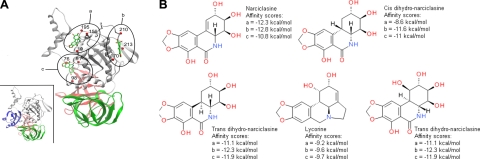
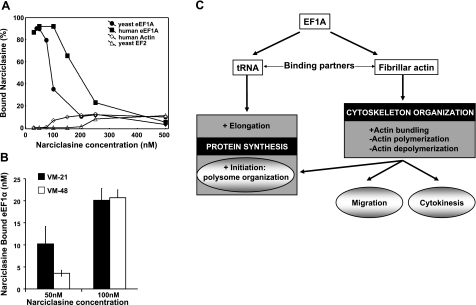
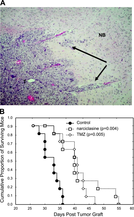
Melanomas display poor response rates to adjuvant therapies because of their intrinsic resistance to proapoptotic stimuli. This study indicates that such resistance can be overcome, at least partly, through the targeting of eEF1A elongation factor with narciclasine, an Amaryllidaceae isocarbostyril controlling plant growth. Narciclasine displays IC(50) growth inhibitory values between 30-100 nM in melanoma cell lines, irrespective of their levels of resistance to proapoptotic stimuli. Normal noncancerous cell lines are much less affected. At nontoxic doses, narciclasine also significantly improves (P=0.004) the survival of mice bearing metastatic apoptosis-resistant melanoma xenografts in their brain. The eEF1A targeting with narciclasine (50 nM) leads to 1) marked actin cytoskeleton disorganization, resulting in cytokinesis impairment, and 2) protein synthesis impairment (elongation and initiation steps), whereas apoptosis is induced at higher doses only (≥200 nM). In addition to molecular docking validation and identification of potential binding sites, we biochemically confirmed that narciclasine directly binds to human recombinant and yeast-purified eEF1A in a nanomolar range, but not to actin or elongation factor 2, and that 5 nM narciclasine is sufficient to impair eEF1A-related actin bundling activity. eEF1A is thus a potential target to combat melanomas regardless of their apoptosis-sensitivity, and this finding reconciles the pleiotropic cytostatic of narciclasine. -


Figures

Figure 1.
Figure 1.
Potential binding sites for the Amaryllidaceae isocarbostyrils on eEF1A protein. A) Three potential binding pockets of narciclasine were found in the 3-D structure of yeast eEF1A in independent docking experiments. They are shown here on the same structure. Pocket a corresponds to the GTP binding site. Pocket c is located in the binding region of the nucleotide exchange factor. The 3 eEF1A domains are colored in gray, red, and green. Positions of residues lining each binding pocket are indicated as red spheres, and their numbers are indicated. Inset: same view of eEF1A structure in association with the nucleotide exchange factor (blue). B) Molecular structures of various AIs and alkaloid lycorine, together with their theoretical affinity scores, calculated for all compounds in each binding site.
Figure 2.
Figure 2.
eEF1As is a target for the AIs. A) Narciclasine binding to eEF1A, actin and EF-2 in vitro: various concentrations of narciclasine were incubated overnight with recombinant yeast eEF1A or human eEF1A isoform 2, human actin, and yeast eEF-2. Bound fraction of narciclasine (expressed as percentage) is calculated on the basis of free narciclasine measured by fluorescence. B) Narciclasine binding to eEF1A in mammalian melanoma cells. C) Summary of the various roles of eEF1A in protein synthesis and actin cytoskeleton organization.
Figure 3.
Figure 3.
Narciclasine-induced effects on actin cytoskeleton and protein synthesis. A) Right-angle light scattering was measured for 1.5 μM of preassembled F-actin. Then 0.5 μM rabbit eEF1A was added in the presence of 0–50 nM narciclasine, and scattering was measured for another 400 s. a, b) Representative graphs at 0 nM (a) and 20 nM narciclasine (b). Asterisk indicates emission shutter closed, eEF1A ± narciclasine added. c) Increased fluorescence intensity on addition of eEF1A to F-actin is reduced in the presence of narciclasine. B) Narciclasine-induced effects on the actin cytoskeleton of VM-21 and VM-48 human melanoma cells treated with 50 nM (IC50 value) narciclasine for 15 min and 3 h, respectively, highlighted by fluorescence. Fibrillar actin is represented in green and globular actin in red. C, E) Spectrometry analysis of polyribosome status in VM-21 (C) and VM-48 (E) human melanoma cells treated with 0 nM (solid circles), 50 nM (open circles), and 100 nM narciclasine (open squares) for 1 h (C) and 5 h (E), of cell extracts centrifuged in sucrose gradients and separated into 33 fractions. D, F) Protein synthesis in VM-21 (D) and VM-48 (F) human melanoma cells left untreated or treated with 50 or 100 nM of narciclasine for 5 and 24 h. After treatment, cells were incubated with a methionine analog that was incorporated in nascent proteins and further biotinylated. Lane 1: cells incubated with the methionine analog alone (negative control). Lanes 2–7: cells incubated for 5 h with the methionine analog. Lane 2: cells treated with 100 μg/ml puromycin for 2 h (positive control). Lane 3: untreated cells. Lanes 4 and 5: cells treated for 5 h with 50 nM and 100 nM narciclasine, respectively. Lanes 6 and 7: cells treated for 24 h with 50 nM and 100 nM narciclasine, respectively. Bottom panels correspond to the Coomassie blue staining of the membranes assessing equal total (labeled and nonlabeled) protein loading and integrity.
Figure 4.
Figure 4.
AI-induced effects on melanoma cell death and proliferation. A) Mean IC50 values obtained with 5 cell lines for 4 AIs by means of the colorimetric MTT test. B) Percentage of cells in a late apoptotic status (double-positive cells for propidium and annexin V stainings analyzed by flow cytometry) when treated with various concentrations of narciclasine. C, E) Flow cytometric analysis of JC-1 staining. Open columns represent green fluorescence of the dye; solid columns represent red fluorescence. D) Western blot analysis of PARP cleavage: PARP full protein (116 kDa) is cleaved during apoptosis in fragments of 85 kDa, while necrotic fragments are of 55 kDa. Tubulin blots assess equal loading and protein integrity. F) Mitosis number per cell over a 72-h period was counted in 5 melanoma cell lines by the videomicroscopic device. Solid bars, control; open bars, 50 nM narciclasine. Results are expressed as means ± se.
Figure 5.
Figure 5.
Narciclasine improves the survival of mice bearing brain melanoma metastatic xenografts. A) Typical hematoxylin eosin staining of a tumor that developed in the brain of a nude mouse 1 mo after the stereotactical graft of human melanoma brain metastatic cells (×50). T, tumor; NB, normal brain tissue; black arrows indicate invasive islets). B) Kaplan-Meier survival analysis of the tumor-bearing mice left untreated, treated with narciclasine (1 mg/kg p.o.; 2 administrations/wk for 3 wk) or with temozolomide (TMZ, 40 mg/kg p.o.; 3 administrations/wk for 3 wk).
Figure 6.
Figure 6.
Correlations of the differential cellular sensitivities in the NCI 60 cell line screen using the COMPARE algorithm: compare correlation coefficients (CCCs) were generated by a computerized pattern-recognition algorithm and serve as an indication of similarities in differential cellular sensitivities or characteristic fingerprints for each compound. Pancratistatin (circles), trans-dihydronarciclasine (triangles), 7-deoxynarciclasine (squares), cis-dihydronarciclasine (large diamonds), and paclitaxel (small diamonds) were each used as a seed to find significant correlations with the anticancer agents in the NCI Standard Compound Database, containing pancratistatin as a representative of the AIs, at the GI50, TGI, and LC50 levels (for definitions of these parameters, see DTP human tumor cell line screen; http://dtp.nci.nih.gov/branches/btb/ivclsp.html). At the GI50 level, all correlations, with the exception of paclitaxel identified pancratistatin, ranked first among all the compounds in the database with CCC > 7 (open markers). Correlations with pancratistatin at the TGI (shaded markers) and LC50 levels (solid markers) were significantly inferior and virtually nonexistent, respectively (CCC 0.4–0.5 and 0.1–0.2). Correlations of paclitaxel with pancratistatin were poor and similar to the rest of the seed compounds at the LC50 level.

Similar articles

Cited by

References

    1. Hamm C., Verma S., Petrella T., Bak K., Charette M. (2008) Melanoma Disease Site Group of Cancer Care Ontario's Program in evidence-based care. Biochemotherapy for the treatment of metastatic malignant melanoma: a systematic review. Cancer Treat. Rev. 34, 145– 146 - PubMed
    1. Atallah E., Flaherty L. (2005) Treatment of metastatic malignant melanoma. Curr. Treat. Options Oncol. 6, 185– 193 - PubMed
    1. Soengas M. S., Lowe S. W. (2003) Apoptosis and melanoma chemoresistance. Oncogene 22, 3138– 3151 - PubMed
    1. Eberle J., Kurbanov B. M., Hossini A. M., Trefzer U., Fecker L. F. (2007) Overcoming apoptosis deficiency of melanoma-hope for new therapeutic approaches. Drug Resist. Updat. 10, 218– 234 - PubMed
    1. Ivanov V. N., Bhoumik A., Ronai Z. (2003) Death receptors and melanoma resistance to apoptosis. Oncogene 22, 3152– 3161 - PubMed

Publication types

MeSH terms