. 2010 Aug 3;107(31):13924-9.
doi: 10.1073/pnas.1000909107. Epub 2010 Jul 19.

NMDA receptor-dependent GABAB receptor internalization via CaMKII phosphorylation of serine 867 in GABAB1

Affiliations

NMDA receptor-dependent GABAB receptor internalization via CaMKII phosphorylation of serine 867 in GABAB1

Nicole Guetg et al. Proc Natl Acad Sci U S A. .

Abstract

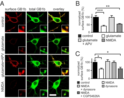
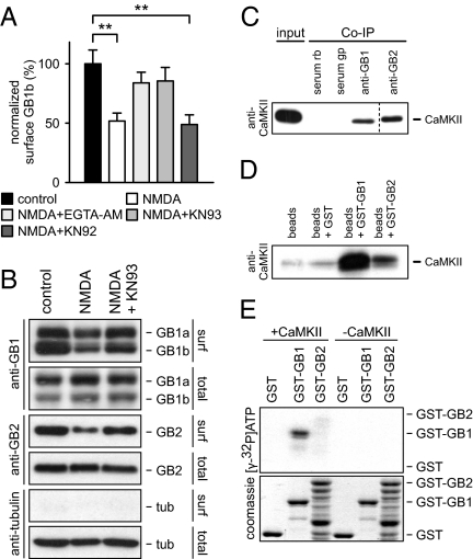
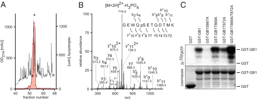
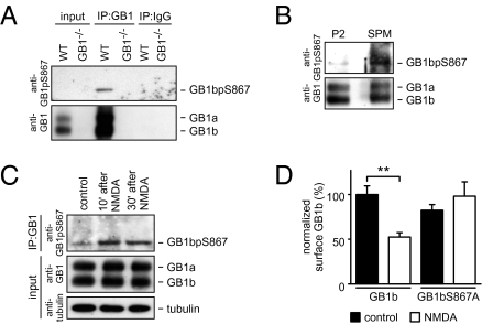
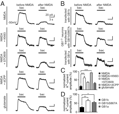
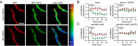
GABAB receptors are the G-protein-coupled receptors for GABA, the main inhibitory neurotransmitter in the brain. GABAB receptors are abundant on dendritic spines, where they dampen postsynaptic excitability and inhibit Ca2+ influx through NMDA receptors when activated by spillover of GABA from neighboring GABAergic terminals. Here, we show that an excitatory signaling cascade enables spines to counteract this GABAB-mediated inhibition. We found that NMDA application to cultured hippocampal neurons promotes dynamin-dependent endocytosis of GABAB receptors. NMDA-dependent internalization of GABAB receptors requires activation of Ca2+/Calmodulin-dependent protein kinase II (CaMKII), which associates with GABAB receptors in vivo and phosphorylates serine 867 (S867) in the intracellular C terminus of the GABAB1 subunit. Blockade of either CaMKII or phosphorylation of S867 renders GABAB receptors refractory to NMDA-mediated internalization. Time-lapse two-photon imaging of organotypic hippocampal slices reveals that activation of NMDA receptors removes GABAB receptors within minutes from the surface of dendritic spines and shafts. NMDA-dependent S867 phosphorylation and internalization is predominantly detectable with the GABAB1b subunit isoform, which is the isoform that clusters with inhibitory effector K+ channels in the spines. Consistent with this, NMDA receptor activation in neurons impairs the ability of GABAB receptors to activate K+ channels. Thus, our data support that NMDA receptor activity endocytoses postsynaptic GABAB receptors through CaMKII-mediated phosphorylation of S867. This provides a means to spare NMDA receptors at individual glutamatergic synapses from reciprocal inhibition through GABAB receptors.


Conflict of interest statement

The authors declare no conflict of interest.

Figures

Fig. 1.
Fig. 1.
NMDA-dependent removal of surface GABAB receptors. (A) Rat hippocampal neurons coexpressing exogenous HA-GB1b-eGFP and GABAB2 were treated at DIV14 as indicated. Surface GABAB1b (GB1b) protein was fluorescence-labeled with anti-HA antibodies before permeabilization. Total GB1b protein was fluorescence-labeled with anti-eGFP antibodies after permeabilization. Single optical planes captured with a confocal microscope are shown. (Scale bar, 15 μm.) (Insets) Representative spines at higher magnification. (B) Surface GABAB1b protein was quantified by the ratio of surface to total fluorescence intensity. Values were normalized to control values in the absence of any pharmacological treatment. Surface GABAB1b protein was significantly decreased following glutamate or NMDA treatment. No significant reduction was observed with glutamate treatment after preincubation with APV. n = 9–10, **P < 0.01, ***P < 0.001. (C) Dynasore but not CGP54626A prevented the NMDA-induced reduction of surface GABAB1b protein. n = 8–10, *P < 0.05. Quantification was from nonsaturated images. Data are presented as mean ± SEM.
Fig. 2.
Fig. 2.
NMDA-induced removal of surface GABAB receptors requires CaMKII. (A) Rat hippocampal neurons coexpressing exogenous HA-GB1b-eGFP and GABAB2 were analyzed at DIV14. Surface GABAB1b protein was quantified by the ratio of surface to total fluorescence intensity. Preincubation of neurons with the Ca2+-chelator EGTA-AM or the CaMKII inhibitor KN-93 prevented the NMDA-induced reduction in surface GABAB1b protein. KN-92 was ineffective. Data are means ± SEM, n = 9–10, **P < 0.01. (B) NMDA-mediated removal of endogenous surface GABAB receptors. Live cortical neurons were treated at DIV14 as indicated and then biotinylated. Cell homogenates (total) and avidin-purified cell surface proteins (surf) were probed on Western blots with anti-GABAB1 (anti-GB1) and anti-GABAB2 (anti-GB2) antibodies. While all GABAB subunits were removed from the cell surface in response to NMDA, GABAB1b was more efficiently removed than GABAB1a (P < 0.05). NMDA-mediated removal of surface protein was inhibited by KN-93. Anti-tubulin antibodies were used as a control. Of note, we consistently observed that significantly more GABAB1b protein was detected at the cell surface under control conditions, albeit GABAB1a is more abundant in cortical neurons (GB1a-to-GB1b ratio: surface, 0.71 ± 0.08; total, 1.32 ± 0.05; n = 3, P < 0.01). (C) CaMKII interacts with GABAB receptors in the brain. Anti-GB1 and anti-GB2 antibodies coimmunoprecipitated CaMKII from purified mouse brain membranes, whereas control rabbit (serum rb) or guinea-pig serum (serum gp) did not. (D) Pull-down assays with GST-fusion proteins containing the entire C-terminal domain of GABAB1 (GST-GB1) or GABAB2 (GST-GB2) and whole-brain lysates. CaMKII bound to a larger extent to GST-GB1 than to GST-GB2. Control assays were with glutathione beads alone or with beads together with GST protein. (E) In vitro phosphorylation of GST-fusion proteins with [γ-32P]-ATP in the presence or absence of recombinant CaMKII. Phosphorylated proteins were separated by SDS/PAGE and exposed to autoradiography. CaMKII specifically phosphorylated GST-GB1 but not GST-GB2 or GST alone. Coomassie blue staining controlled for loading. The GST-GB2 fusion protein tended to degrade (50).
Fig. 3.
Fig. 3.
CaMKII phosphorylates S867 in the GABAB1 subunit. (A) RP-HPLC analysis of proteolytically digested GST-GB1 after phosphorylation with recombinant CaMKII and [γ-32P]-ATP. Peptide elution was monitored at 214 nm and radioactivity (red) determined by liquid scintillation counting. Asterisk marks elution of the 32P-labeled peptide in fraction 54. (B) Fragmentation spectrum of the doubly charged 768.29 Da precursor from the phosphorylated peptide of fraction 54. Fragmentation pattern agrees with predicted ESI-MS/MS spectrum for the phosphopeptide GEWQpS867ETQDTMK. The y- and b-ions matching the GEWQpS867ETQDTMK sequence are labeled. Asterisks mark phosphorylated ions. (C) In vitro phosphorylation of GST fusion proteins with recombinant CaMKII and [γ-32P]-ATP. Phosphorylated proteins were separated by SDS/PAGE and exposed to autoradiography. Substitution of S867 with alanine in GST-GB1S867A prevented phosphorylation by CaMKII, whereas alanine substitutions of other putative phosphorylation sites in proximity of S867 (GST-GB1T869A, GST-GB1T872A and GST-GB1T869A/T872A) did not. Coomassie blue staining controlled for loading.
Fig. 4.
Fig. 4.
S867 phosphorylation in brain tissue and cultured neurons. (A) S867 phosphorylation was detectable after immunoprecipitation of GABAB receptors with anti-GABAB1 antibodies (IP:GB1) from WT but not GB1−/− brain membranes. S867 phosphorylation was detected on Western blots with a phosphorylation-state specific antibody (anti-GB1pS867). The same blot was reprobed with anti-GB1 antibodies. Immunoprecipitation with rabbit IgG (IP:IgG) was used as a control. Note the specific phosphorylation of the GABAB1b subunit. (B) S867 phosphorylation of GABAB1b was clearly detectable in synaptic plasma membranes (SPM) and barely detectable in the P2 membrane fraction purified from total mouse brain homogenates. (C) NMDA application to cultured cortical neurons increased S867 phosphorylation in the GABAB1b subunit. Neurons were treated with NMDA for 3 min and harvested at the times indicated. Whole-cell lysates (input) were subjected to immunoprecipitation with anti-GB1 antibodies (IP:GB1). S867 phosphorylation was detected on Western blot with anti-GB1pS867; anti-tubulin antibodies were used as control. (D) Alanine mutation of S867 in GABAB1b prevents NMDA-induced internalization. Cultured hippocampal neurons expressing exogenous HA-GB1b-eGFP (GB1b) or HA-GB1bS867A-eGFP (GB1bS867A) together with GABAB2 were analyzed at DIV14. Surface GABAB1b protein was quantified by the ratio of surface to total fluorescence intensity. Values were normalized to GB1b control in the absence of NMDA. Data are means ± SEM, n = 8–10. **P < 0.01.
Fig. 5.
Fig. 5.
CaMKII reduces GABAB-mediated K+ currents in cultured hippocampal neurons. (A) Representative baclofen-induced K+ currents recorded at −50 mV before and after application of NMDA or glutamate. Baclofen-induced K+ currents were strongly reduced 30 min after NMDA or glutamate application. KN-93 and dCPP but not STO-609 attenuated the NMDA-mediated K+ current reduction. (B) Representative baclofen-induced K+ currents recorded from neurons of GABAB1−/− (GB1−/−) mice transfected with GABAB1a (GB1a) GABAB1b, (GB1b) or GB1bS867A expression vectors. NMDA was less effective in decreasing the K+ current in neurons transfected with GB1bS867A or GABAB1a. (C) Bar graph illustrating that dCPP and KN-93 attenuated the NMDA-mediated reduction of baclofen-induced K+ currents. (D) NMDA was significantly less effective in decreasing the K+ current in GB1−/− neurons transfected with GB1a or GB1bS867A than with GB1b. Maximal K+-current amplitudes after NMDA application were normalized to the maximal K+-current amplitudes before NMDA application. Data are means ± SEM, n = 3–6. *P < 0.05; **P < 0.01; ***P < 0.001.
Fig. 6.
Fig. 6.
NMDA-mediated endocytosis of GABAB receptors in dendritic spines and shafts. (A) Red fluorescence (R), green fluorescence (G), and G/R ratio images of dendrites expressing freely diffusible RFP and SEP-GB1b before and after NMDA application. NMDA application leads to a decrease in green fluorescence in dendritic spines and shafts. G/R ratio is coded in rainbow colors and is scaled to encompass 2 SDs (2σ) of the average dendritic ratio before NMDA application. (Scale bar, 5 μm.) (B and C) Time course of red and green fluorescence in dendritic spines (B) and shafts (C) before and after NMDA application. NMDA leads to a long-lasting decrease in SEP-fluorescence within minutes, which is prevented by prior application of dCPP. Data are mean ± SEM.

Similar articles

Cited by

References

    1. Bettler B, Kaupmann K, Mosbacher J, Gassmann M. Molecular structure and physiological functions of GABAB receptors. Physiol Rev. 2004;84:835–867. - PubMed
    1. Couve A, Moss SJ, Pangalos MN. GABAB receptors: A new paradigm in G protein signaling. Mol Cell Neurosci. 2000;16:296–312. - PubMed
    1. Ulrich D, Bettler B. GABAB receptors: Synaptic functions and mechanisms of diversity. Curr Opin Neurobiol. 2007;17:298–303. - PubMed
    1. Kornau HC. GABAB receptors and synaptic modulation. Cell Tissue Res. 2006;326:517–533. - PubMed
    1. Jarolimek W, Misgeld U. GABAB receptor-mediated inhibition of tetrodotoxin-resistant GABA release in rodent hippocampal CA1 pyramidal cells. J Neurosci. 1997;17:1025–1032. - PMC - PubMed

Publication types

Substances