. 2010 Aug 3;107(31):13724-9.
doi: 10.1073/pnas.1008117107. Epub 2010 Jul 19.

Aggregation of human mesenchymal stromal cells (MSCs) into 3D spheroids enhances their antiinflammatory properties

Affiliations

Aggregation of human mesenchymal stromal cells (MSCs) into 3D spheroids enhances their antiinflammatory properties

Thomas J Bartosh et al. Proc Natl Acad Sci U S A. .

Abstract

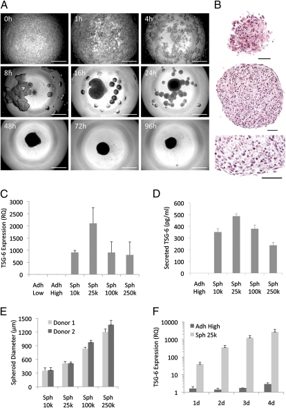
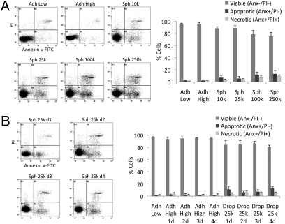
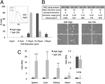
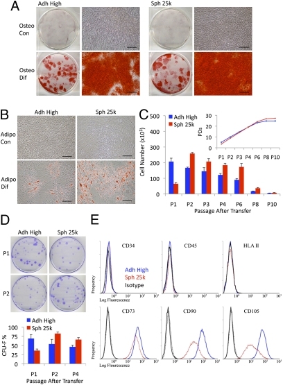
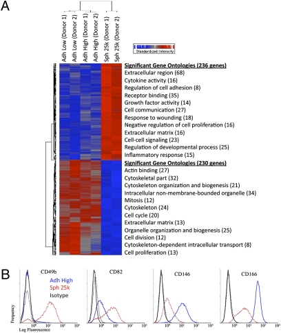
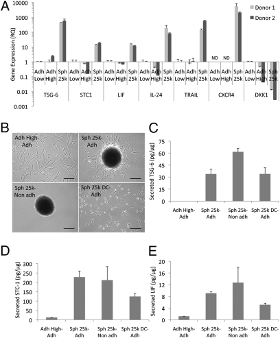
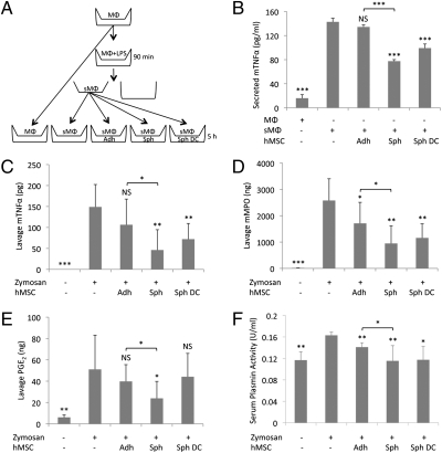
Previous reports suggested that culture as 3D aggregates or as spheroids can increase the therapeutic potential of the adult stem/progenitor cells referred to as mesenchymal stem cells or multipotent mesenchymal stromal cells (MSCs). Here we used a hanging drop protocol to prepare human MSCs (hMSCs) as spheroids that maximally expressed TNFalpha stimulated gene/protein 6 (TSG-6), the antiinflammatory protein that was expressed at high levels by hMSCs trapped in the lung after i.v. infusion and that largely explained the beneficial effects of hMSCs in mice with myocardial infarcts. The properties of spheroid hMSCs were found to depend critically on the culture conditions. Under optimal conditions for expression of TSG-6, the hMSCs also expressed high levels of stanniocalcin-1, a protein with both antiinflammatory and antiapoptotic properties. In addition, they expressed high levels of three anticancer proteins: IL-24, TNFalpha-related apoptosis inducing ligand, and CD82. The spheroid hMSCs were more effective than hMSCs from adherent monolayer cultures in suppressing inflammatory responses in a coculture system with LPS-activated macrophages and in a mouse model for peritonitis. In addition, the spheroid hMSCs were about one-fourth the volume of hMSCs from adherent cultures. Apparently as a result, larger numbers of the cells trafficked through the lung after i.v. infusion and were recovered in spleen, liver, kidney, and heart. The data suggest that spheroid hMSCs may be more effective than hMSCs from adherent cultures in therapies for diseases characterized by sterile tissue injury and unresolved inflammation and for some cancers that are sensitive to antiinflammatory agents.


Conflict of interest statement

The authors declare no conflict of interest.

Figures

Fig. 1.
Fig. 1.
The expression of TSG-6 was increased as hMSCs aggregated into spheroids in hanging drops. (A) Phase contrast microscopy showing the time course of the aggregation of 25,000 hMSCs into a spheroid in a hanging drop. (Scale bar, 500 μm.) (B) H&E staining of hMSC spheroid sections from 3-d hanging drop cultures. Surface (Top), and center (Middle and Bottom) of a spheroid. (Scale bar, 50 μm.) (C) Real-time RT PCR measurements of TSG-6 expression in hMSCs shown as relative to Adh Low sample (n = 3). (D) ELISA measurements of TSG-6 secretion over 24 h from hMSCs grown for 3 d at high density or as hanging drops at different cell densities (n = 4). (E) Sizes of spheroids generated by hMSCs from two donors grown in hanging drops for 3 d. Sizes were measured from captured images of transferred spheroids (n = 7–13). (F) Real-time RT PCR measurements of TSG-6 expression in hMSCs grown at high density or in hanging drops at 25,000 cells/drop for 1–4 d shown as relative to hMSCs grown at low density (n = 3). Values are mean ± SD. Abbreviations: RQ, relative quantity; Adh Low, hMSCs plated at 100 cells/cm2 for 7–8 d until about 70% confluent; Adh High, hMSCs harvested from same Adh Low cultures, plated at 5,000 cells/cm2 and incubated for 3 d; Sph 10k-250k, hMSCs harvested from same Adh Low cultures and incubated for 3 d in hanging drops at 10,000-250,000 cells/drop.
Fig. 2.
Fig. 2.
Viability of hMSCs in spheroids. (A and B) Viability of hMSCs as determined by flow cytometry measuring PI uptake and annexin V-FITC labeling. Spheroids were dissociated with trypsin/EDTA. Representative log fluorescent dot plots and summary of the data are shown. Values are mean ± SD (n = 3). Abbreviations: As in Fig. 1 with 1d to 4d indicating days of incubation.
Fig. 3.
Fig. 3.
Size analysis and i.v. infusion of spheroid hMSCs. (A) Assays of cell size by flow cytometry (n = 3). hMSC sizes were estimated from forward scatter (FS) (Inset) properties of the viable population (calcein AM+/7AAD) relative to beads with known diameters (3, 7, 15, and 25 μm). (B) Cell size assayed by microscopy. (C) Relative tissue distribution of i.v. infused hMSCs. NOD/scid mice were infused i.v. with 106 monolayer or spheroid derived hMSCs. After 15 min, tissues were harvested for genomic DNA and tissue distribution of hMSCs was determined with real-time PCR for human Alu and GAPDH (n = 4–5) and shown as relative to Adh High sample. *P < 0.05, **P < 0.01, and ***P < 0.001. Values are mean ± SD. Abbreviations: as in Fig. 1.
Fig. 4.
Fig. 4.
Spheroid hMSCs retain the properties of hMSCs from adherent cultures. (A) Differentiation of hMSCs in osteogenic medium (Osteo Dif) and control medium (Osteo Con). Cultures were stained with Alizarin Red after 14 d. (Scale bar, 200 μm.) (B) Differentiation of hMSCs in adipogenic medium (Adipo Dif) and control medium (Adipo Con). Cultures were stained with Oil Red O after 14 d. (Scale bar, 200 μm.) (C) Growth of hMSCs (donor 2) as monolayers from high density and hanging drop cultures plated at low density (5,500 cells/plate) and passaged every 7 d (n = 4). Cumulative population doublings (PDs) after each passage are shown (Inset). (D) CFU-F assays of hMSCs (donor 2) plated at 83 cells/plate and incubated for 14 d (n = 4). Representative plates at passage 1 and passage 2 after transfer. Values are mean ± SD. (E) Flow cytometry of surface protein expression on hMSCs. Abbreviations: as in Fig. 1 with P1 to P10 indicating passage number.
Fig. 5.
Fig. 5.
Microarray assays of hMSCs from two donors. (A) Hierarchical clustering of differentially expressed genes. Genes that were either up- (236 genes) or down-regulated (230 genes) in spheroids (Sph 25k) at least twofold compared with their adherent culture counterparts (Adh Low and Adh High), were used in hierarchical clustering. The most significant Gene Ontology terms for up-regulated genes (red) and down-regulated genes (blue) are shown next to the heat map. (B) Flow cytometry of differentially expressed surface epitopes on hMSCs. Abbreviations: as in Fig. 1.
Fig. 6.
Fig. 6.
Spheroid hMSCs express high levels of antiinflammatory and antitumorigenic molecules. (A) Real-time RT PCR measurements for antiinflammatory genes (TSG-6, STC-1, and LIF), antitumorigenic genes (IL-24 and TRAIL), gene for an MSC homing receptor (CXCR4), and gene for the Wnt signaling inhibitor (DKK1) for two donors. Values are mean RQ ± 95% confidence interval from triplicate assays compared with Adh Low sample. (B) Images of high density monolayer (Adh High), spheroids (Sph 25k), and spheroid derived hMSCs (Sph 25k DC) 24 h after transfer onto adherent (Adh) or nonadherent (Non adh) surfaces. Cultures were in six-well plates containing 1.5 mL CCM and either 200,000 hMSCs from high density cultures, eight spheroids, or 200,000 hMSCs dissociated from spheroids. After 24 h, medium was recovered for ELISAs and cells lyzed for protein assays. (Scale bar, 200 μm.) TSG-6 (C), STC-1 (D), and LIF (E) ELISAs on medium, normalized to total cellular protein. Values are mean ± SD (n = 3). Abbreviations: as in Fig. 1 with ND indicating not detectable and Sph 25k DC-Adh indicating hMSCs dissociated from Sph 25k and plated on cell adherent surfaces.
Fig. 7.
Fig. 7.
hMSC spheroids exhibit enhanced antiinflammatory effects in vitro and in vivo. (A) Schematic of the mouse macrophage (mMΦ) assay. mMΦs were seeded in the upper chamber of a transwell, stimulated with LPS for 90 min, the LPS was removed, and the chamber transferred to a six-well dish plated with monolayer (Adh), spheroid (Sph), or spheroid-derived hMSCs (Sph DC) at the same cell density. MΦ:hMSC (2:1). After 5 h, medium was collected for ELISAs. (B) ELISA for mTNFα in medium from cocultures (n = 3). (CF) Antiinflammatory activity of hMSCs in a mouse model of peritonitis. C57BL/6 mice were injected i.p. with zymosan to induce inflammation. After 15 min, the mice were injected i.p. with 1.5 × 10⁶ monolayer hMSCs, 60 spheroids, or 1.5 × 10⁶ spheroid derived cells. After 6 h, peritoneal lavage was collected and mTNFα (C), mMPO (D), and PGE2 (E) levels were determined with ELISAs. Total amounts of the specific molecules in the lavage are shown (n = 4–8). After 24 h, blood was collected and plasmin activity was measured from serum (n = 3–6). Values are mean ± SD. Not significant (NS) P ≥ 0.05, *P < 0.05, **P < 0.01, and ***P < 0.001. Abbreviations: as in Figs. 1 and 6.

Similar articles

Cited by

References

    1. Friedenstein AJ, Gorskaja JF, Kulagina NN. Fibroblast precursors in normal and irradiated mouse hematopoietic organs. Exp Hematol. 1976;4:267–274. - PubMed
    1. Owen M, Friedenstein AJ. Stromal stem cells: Marrow-derived osteogenic precursors. Ciba Found Symp. 1988;136:42–60. - PubMed
    1. Caplan AI. Mesenchymal stem cells. J Orthop Res. 1991;9:641–650. - PubMed
    1. Eaves CJ, et al. Molecular analysis of primitive hematopoietic cell proliferation control mechanisms. Ann N Y Acad Sci. 1991;628:298–306. - PubMed
    1. Dominici M, et al. Minimal criteria for defining multipotent mesenchymal stromal cells. The International Society for Cellular Therapy position statement. Cytotherapy. 2006;8:315–317. - PubMed

Publication types

MeSH terms

LinkOut - more resources