. 2010 Aug 3;107(31):13848-53.
doi: 10.1073/pnas.1003020107. Epub 2010 Jul 19.

Alterations in the immuno-skeletal interface drive bone destruction in HIV-1 transgenic rats

Affiliations

Alterations in the immuno-skeletal interface drive bone destruction in HIV-1 transgenic rats

Tatyana Vikulina et al. Proc Natl Acad Sci U S A. .

Abstract

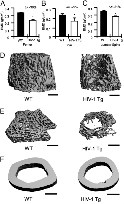
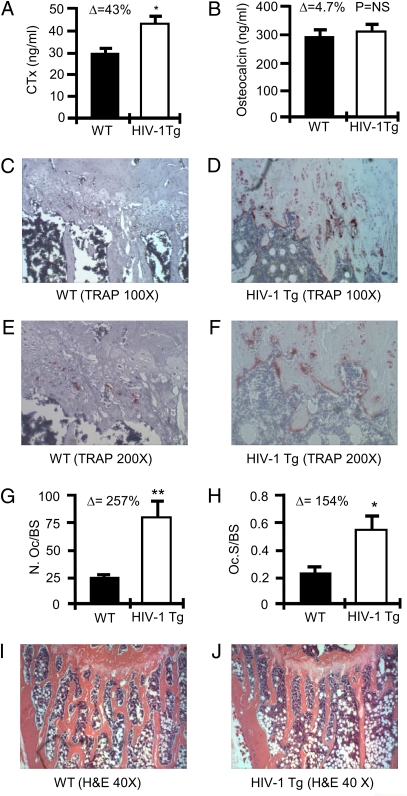
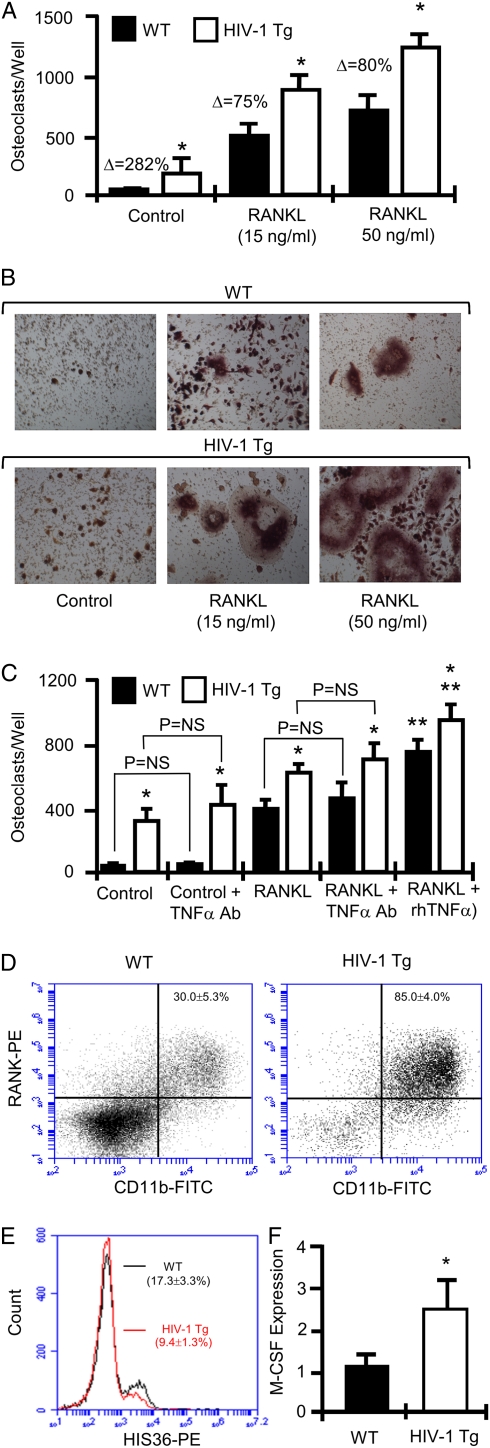
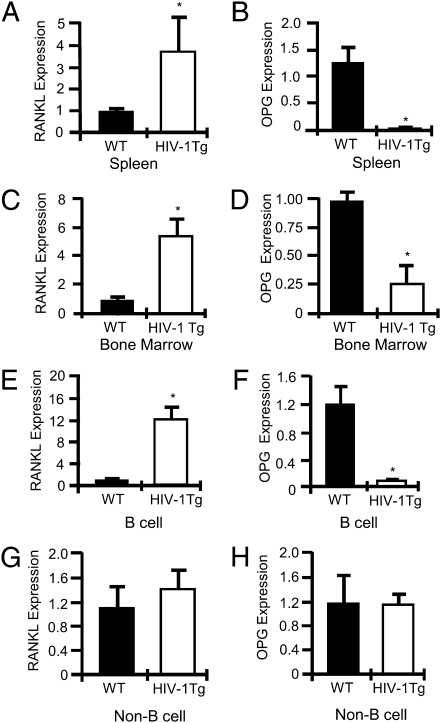
Osteoporosis and bone fractures are increasingly recognized complications of HIV-1 infection. Although antiretroviral therapy itself has complex effects on bone turnover, it is now evident that the majority of HIV-infected individuals already exhibit reduced bone mineral density before therapy. The mechanisms responsible are likely multifactorial and have been difficult to delineate in humans. The HIV-1 transgenic rat recapitulates many key features of human AIDS. We now demonstrate that, like their human counterparts, HIV-1 transgenic rats undergo severe osteoclastic bone resorption, a consequence of an imbalance in the ratio of receptor activator of NF-kappaB ligand, the key osteoclastogenic cytokine, to that of its physiological decoy receptor osteoprotegerin. This imbalance stemmed from a switch in production of osteoprotegerin to that of receptor activator of NF-kappaB ligand by B cells, and was further compounded by a significantly elevated number of osteoclast precursors. With the advancing age of individuals living with HIV/AIDS, low bone mineral density associated with HIV infection is likely to collide with the pathophysiology of skeletal aging, leading to increased fracture risk. Understanding the mechanisms driving bone loss in HIV-infected individuals will be critical to developing effective therapeutic strategies.


Conflict of interest statement

The authors declare no conflict of interest.

Figures

Fig. 1.
Fig. 1.
BMD and bone structure in HIV-1 Tg rats. BMD in (A) femurs, (B) tibias, and (C) lumbar spines were analyzed by DXA ex vivo. The percentage change (Δ) between WT and HIV-1 Tg is indicated for each site. (n = 4 femurs or tibias per group and n = 6 spines per group; average ± SD, *P ≤ 0.05 by Mann-Whitney test). (D) Representative longitudinal trabecular (12 μm), (E) cross-sectional (6 μm) trabecular, and (F) cortical (12 μm) 3D reconstructions of femurs from WT and HIV-1 Tg rats were generated by μCT. (Scale bar, 1 mm.)
Fig. 2.
Fig. 2.
OCs and bone resorption in HIV-1 Tg rats. (A) C-terminal telopeptide (CTx) and (B) osteocalcin were quantified in the serum of WT and HIV Tg rats. (*P ≤ 0.002 by t test; n = 4 rats per group). Histology and histomorphometry for OCs were performed in tibias. Representative TRAP-stained tibia sections are shown for WT rats (C and E) and HIV-1 Tg rats (D and F) at magnifications of 100× (C and D) and 200× (E and F). (G) The number of OCs per bone surface area (N.Oc/BS); and (H) OC surface per bone surface area (Oc.S/BS) was calculated as mean ± SD of five WT and six HIV-1 Tg rats (*P = 0.03, **P = 0.0095 by t test). H&E-stained tibias for WT (I) and HIV-1 Tg (J) rats at 40× magnification. Mineralized bone stains orange/pink.
Fig. 3.
Fig. 3.
In vitro OC formation and quantification of OC precursors. (A) OCs were cultured from BM isolated from WT and HIV-1 Tg rats in the absence of RANKL (control), subsaturating concentrations of RANKL (15 ng/mL), and saturating concentrations of RANKL (50 ng/mL). TRAP positive multinucleated cells were scored as OCs. Mean ± SD of three independent experiments with six wells per data set each. (n = 2 rats/group pooled for each experiment; *P < 0.01 on one-way ANOVA with Tukey-Kramer post-test; Δ, percentage change from WT.) (B) Photomicrographs of representative OC cultures. (C) OCs were cultured from BM isolated from WT and HIV-1 Tg rats in the presence or absence of RANKL (15 ng/mL) and neutralizing antibody to TNF-α (TNF-α Ab; 20 μg/mL) or rmTNF-α (2.5 ng/mL). TRAP-positive multinucleated cells (≥3 nuclei) were scored as OCs. Mean ± SD of two independent experiments of six wells per data set each. (n = 1 rat per group for each experiment; *P < 0.001 vs. WT, **P < 0.001 vs. RANKL only, P = not significant, one-way ANOVA with Tukey-Kramer post test.) (D) FACS analysis of OC precursors (CD11b+RANK+ cells) and (E) HIS36+ macrophages. (F) Real-time RT-PCR for whole BM M-CSF expression. (Mean ± SD of five rats per group assayed independently; *P = 0.032, Mann-Whitney test.)
Fig. 4.
Fig. 4.
Total and B cell RANKL and OPG expression in HIV-1 Tg rat spleen and BM. Real-time RT-PCR for RANKL in whole spleen (A) and BM (C), and for OPG in whole spleen (B) and BM (D). RANKL (E) and OPG (F) expression in purified splenic B cells. RANKL (G) and OPG (H) expression by B cell–depleted splenocytes. (*P < 0.002 by t test, n = 4 rats per group.)

References

    1. Li Y, et al. B cells and T cells are critical for the preservation of bone homeostasis and attainment of peak bone mass in vivo. Blood. 2007;109:3839–3848. - PMC - PubMed
    1. Weitzmann MN, Pacifici R. Estrogen deficiency and bone loss: An inflammatory tale. J Clin Invest. 2006;116:1186–1194. - PMC - PubMed
    1. Eghbali-Fatourechi G, et al. Role of RANK ligand in mediating increased bone resorption in early postmenopausal women. J Clin Invest. 2003;111:1221–1230. - PMC - PubMed
    1. Deeks SG. Immune dysfunction, inflammation, and accelerated aging in patients on antiretroviral therapy. Top HIV Med. 2009;17:118–123. - PubMed
    1. Nguyen N, Holodniy M. HIV infection in the elderly. Clin Interv Aging. 2008;3:453–472. - PMC - PubMed

Publication types

LinkOut - more resources