

Low-dose, simple, and fast grating-based X-ray phase-contrast imaging
- PMID: 20643971
- PMCID: PMC2922255
- DOI: 10.1073/pnas.1003198107
Low-dose, simple, and fast grating-based X-ray phase-contrast imaging
Abstract
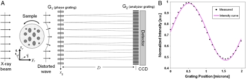
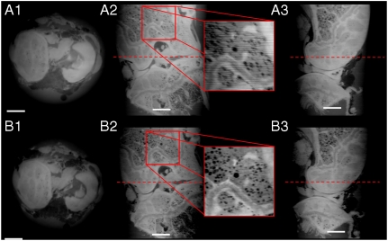
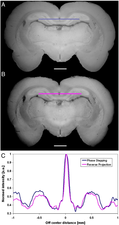
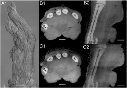
Phase sensitive X-ray imaging methods can provide substantially increased contrast over conventional absorption-based imaging and therefore new and otherwise inaccessible information. The use of gratings as optical elements in hard X-ray phase imaging overcomes some of the problems that have impaired the wider use of phase contrast in X-ray radiography and tomography. So far, to separate the phase information from other contributions detected with a grating interferometer, a phase-stepping approach has been considered, which implies the acquisition of multiple radiographic projections. Here we present an innovative, highly sensitive X-ray tomographic phase-contrast imaging approach based on grating interferometry, which extracts the phase-contrast signal without the need of phase stepping. Compared to the existing phase-stepping approach, the main advantages of this new method dubbed "reverse projection" are not only the significantly reduced delivered dose, without the degradation of the image quality, but also the much higher efficiency. The new technique sets the prerequisites for future fast and low-dose phase-contrast imaging methods, fundamental for imaging biological specimens and in vivo studies.
Conflict of interest statement
The authors declare no conflict of interest.
Figures
References
-
- Momose A, Fukuda J. Phase-contrast radiographs of nonstained rat cerebellar specimen. Med Phys. 1995;22:375–379. - PubMed
-
- Bonse U, Hart M. An x-ray interferometer with long separated interfering beam paths. Appl Phys Lett. 1965;6:155–156.
-
- Ando M, Hosoya S. In: Shinoda G, Kohra K, Ichinokawa T, editors. Proceedings of the 6th International Conference of X-ray Optics and Microanalysis; Tokyo: Univ of Tokyo Press; 1972. pp. 63–68.
-
- Momose A, Takeda T, Itai Y, Hirano K. Phase-contrast X-ray computed tomography for observing biological soft tissues. Nat Med. 1996;2:473–475. - PubMed
-
- Snigirev A, Snigireva I, Kohn V, Kuznetsov S, Schelokov I. On the possibilities of x-ray phase contrast microimaging by coherent high-energy synchrotron radiation. Rev Sci Instrum. 1995;66:5486–5492.
Publication types
MeSH terms
LinkOut - more resources
Full Text Sources
Other Literature Sources
