. 2010 Sep;56(3):397-404.
doi: 10.1161/HYPERTENSIONAHA.110.153262. Epub 2010 Jul 19.

Estradiol stimulates capillary formation by human endothelial progenitor cells: role of estrogen receptor-{alpha}/{beta}, heme oxygenase 1, and tyrosine kinase

Affiliations

Estradiol stimulates capillary formation by human endothelial progenitor cells: role of estrogen receptor-{alpha}/{beta}, heme oxygenase 1, and tyrosine kinase

Isabella Baruscotti et al. Hypertension. 2010 Sep.

Abstract

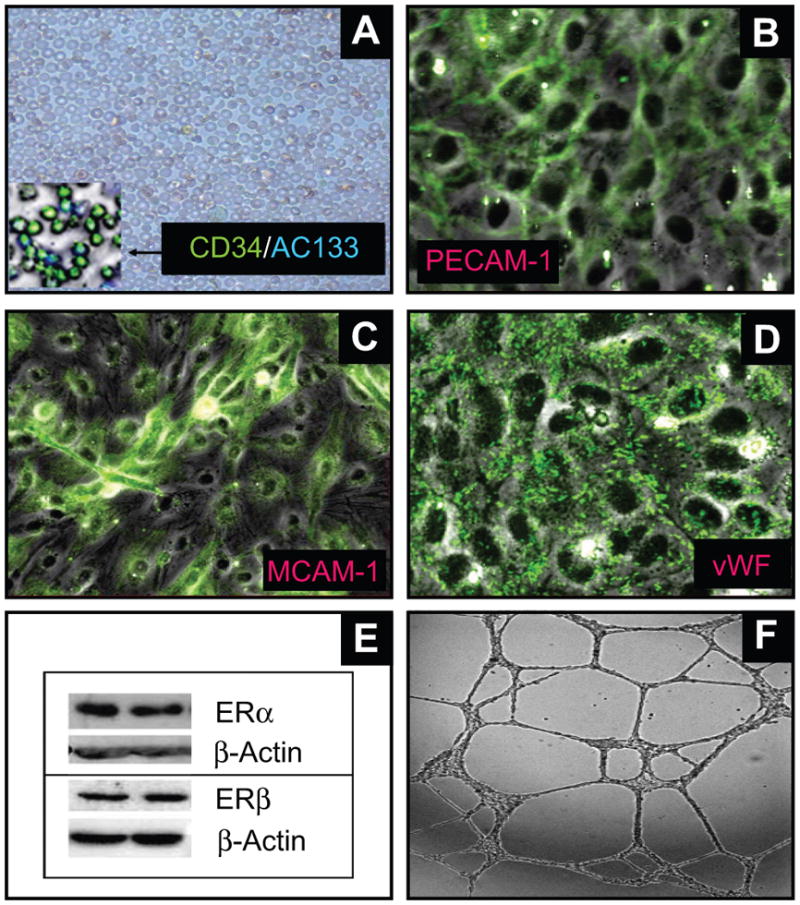
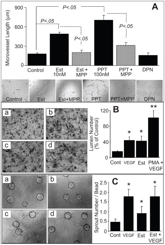
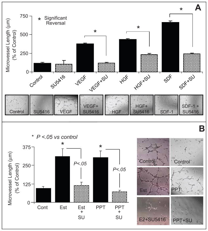
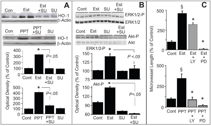
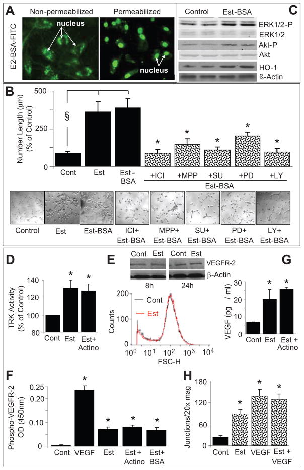
Endothelial progenitor cells (EPCs) repair damaged endothelium and promote capillary formation, processes involving receptor tyrosine kinases (RTKs) and heme oxygenase 1 (HO-1). Because estradiol augments vascular repair, we hypothesize that estradiol increases EPC proliferation and capillary formation via RTK activation and induction of HO-1. Physiological concentrations of estradiol (10 nmol/L) increased EPC-induced capillary sprout and lumen formation in matrigel/fibrin/collagen systems. Propyl-pyrazole-triol (PPT; 100 nmol/L; estrogen receptor [ER]-alpha agonist), but not diarylpropionitrile (ER-beta agonist), mimicked the stimulatory effects of estradiol on capillary formation, and methyl-piperidino-pyrazole (ER-alpha antagonist) abolished the effects of estradiol and PPT. Three different RTK activators (vascular endothelial growth factor, hepatocyte growth factor, and stromal derived growth factor 1) mimicked the capillary-stimulating effects of estradiol and PPT. SU5416 (RTK inhibitor) blocked the stimulatory effects of estradiol and PPT on capillary formation. Estradiol increased HO-1 expression by 2- to 3-fold, an effect blocked by SU5416, and PPT mimicked the effects of estradiol on HO-1. The ability of estradiol to enhance capillary formation, increase expression of HO-1, and augment phosphorylation of extracellular signal-regulated kinase 1/2, Akt, and vascular endothelial growth factor receptor 2 was mimicked by its cell-impermeable analog BSA estradiol. Actinomycin (transcription inhibitor) did not alter the effects of estradiol on RTK activity or vascular endothelial growth factor secretion. We conclude that estradiol via ER-alpha promotes EPC-mediated capillary formation by a mechanism that involves nongenomic activation of RTKs and HO-1 activation. Estradiol in particular and ER-alpha agonists in general may promote healing of injured vascular beds by promoting EPC activity leading to more rapid endothelial recovery and capillary formation after injury.


Figures

Figure 1
Figure 1
Representative photomicrographs and Western blot showing that circulating CD34+ progenitor cells can transform into endothelial cells and express estrogen receptors. (A) depicts the CD34+ cells isolated following antibody based magnetic separation. The inset depicts the purity of the cells stained with CD34 and AC133 antibodies; Panels B-D depicts that progenitor cell-derived endothelial cells express the endothelial cell markers: platelet endothelial cell adhesion molecule-1 (PECAM-1; B); melanoma cell adhesion molecule-1 (MCAM-1; C) and von Willebrand factor (vWF; D). (E) Representative Western blots demonstrating that EPCs express both estrogen receptors (ERs) α and β. Photomicrograph in panel F depicts the functional capability of EPCs to form capillaries in response to serum.
Figure 2
Figure 2
(A) Bar graph and representative photomicrographs showing the effects of estradiol (Est; 10nmol/L), PPT (ER-α agonist; 100nmol/L) and DPN (100nmol/L) on capillary formation by EPCs in the presence and absence of the ER-α antagonist MPP (1μmol/L). Serum starved EPCs were plated at a density of 50,000 cells/200μl/well on matrigel coated 8-well multichamber slides. After 4 hours the capillary formation was assessed by microscopically measuring the length of capillaries at 5 random locations (4x-magnification). Values represent means±SEM. (B) EPC-EC were seeded into collagen gels (3.75mg/ml) in EGM2 medium and incubated for 24 hours as follows: (a) control (Cont); (b) VEGF (40ng/ml); (c) estradiol (Est,10nmol/L); or (d), positive control[phorbol myristate actetate (PMA;50ng/ml) + VEGF]. Cultures were subsequently fixed in 2% paraformaldehyde, stained with toluidine blue and photographed. Shown are mean number of lumens and SD; *(P<0.05), **(P<0.01) relative to control, by Student t test. (C) EPC-EC-coated Cytodex3 beads in fibrin gels were cultured in EGM2 medium as follows: (a) without VEGF; (b) with VEGF (15ng/ml); (c) with estradiol (Est,10nmol/L); or (d) with VEGF+E2. Gels were photographed and the number of sprouts counted after 7 days. Shown are means and SD; *(P<0.05), **(P<0.01) relative to control, by Student t test.
Figure 3
Figure 3
(A) Bar graph and representative photomicrographs showing the effects of receptor tyrosine kinase stimulators vascular endothelial growth factor (VEGF;100ng/mL), hepatocyte growth factor (HGF, 100ng/mL) and stromal cell-derived growth factor (SDF-1 100ng/mL) on capillary formation by EPCs in the presence and absence of the tyrosine kinase inhibitor SU5416 (SU; 5μmol/L). Serum starved EPCs were plated at a density of 50,000 cells/200μl/well on matrigel coated 8-well multichamber slides. After 4 hours the capillary formation was assessed by microscopically measuring the length of capillaries at 5 random locations (4x-magnification). (B) Bar graph and representative photomicrographs showing the modulatory effects of the tyrosine kinase inhibitor SU5416 (SU;5μmol/L) on estradiol (10nmol/L) and PPT (100nmol/L) induced capillary formation in EPCs. Values represent means±SEM. * p<0.05 versus vehicle treated controls.
Figure 4
Figure 4
Bar graph and representative Western blots demonstrating that estradiol induces hemeoxygenase-1 (HO-1) expression in EPCs via estrogen receptor (ER)-α. Panel A depicts the effects of estradiol (Est; 10nmol/L), PPT (ER-α agonist; 100nmol/L) and DPN ( ER-β agonist; 100nmol/L) on HO-1 expression in EPCs treated for 4 hours. Panel B depicts the effects of Est (10nmol/L) and PPT (100nmol/L) on HO-1 expression in EPCs in the presence and absence of the ER-α antagonist MPP (1μmol/L). Cells were pretreated for 15-minutes with MPP and subsequently estradiol or PPT were added for an additional 4 hours. Values represent means±SEM.
Figure 5
Figure 5
(A) Bar graph and representative Western blots demonstrating that inhibition of tyrosine kinase with SU5416 (SU) abrogates the stimulatory effects of estradiol and PPT on HO-1 expression in EPCs. EPCs were pretreated with SU (5μmol/L) for 15 minutes and subsequently estradiol (Est; 10nmol/L) or PPT (100nmol/L) was added for an additional 4 hours. HO-1 expression was assayed in the lysates with β-actin as control. Bargraph depicts the densitometry analysis of the HO-1 bands, which were normalize to β-actin. * p<0.05 versus vehicle treated control. Panel B: Depicts the modulatory effects of SU5416 (SU) on estradiol-induced ERK1/2 and Akt phosphorylation in EPCs. Cells were pretreated with SU for 15 minutes and subsequently estradiol (100nmol/L) was added for an additional 10 minutes. Cell lysates were prepared and expression of phosphorylated ERK1/2 and Akt were analysed by Western blotting. For normalization non-phosphorylated ERK1/2 and Akt were used. Bar graph depicts the densitometry analysis of the phosphorylated ERK1/2 (ERK1/2-P) and Akt (Akt-P) bands, which were normalize to ERK1/2 and Akt, respectively. * p<0.05 versus vehicle treated control. Values represent means±SEM. (C) Bar graph showing the inhibitory effects of LY294002 (Akt inhibitor; LY; 10μmol/L) and PD98059 (ERK1/2-P inhibitor; PD; 10μmol/L) on estradiol (Est; 10nmol/L) and PPT (100nmol/L) induced capillary formation by EPCs. EPCs were pretreated for 15 minutes with either LY or PD and subsequently Est or PPT added for another 4hoursand capillary formation analyzed microscopically at 4x magnifcation. * p<0.05 vs Estradiol or PPT alone.
Figure 6
Figure 6
(A) Representative photomicrographs providing evidence that BSA-estradiol is impermeable and stains the nucleus of permeabilized, but not intact, EPCs (original magnification 20x); (B) Bar graph and representative photomicrographs showing the modulatory effects of receptor tyrosine kinase inhibitor SU5416 (SU; 5μmol/L), LY294002 (Akt pathway inhibitor; LY, 10μmol/L) and PD98059 (ERK1/2 pathway inhibitor; PD, 10 μmol/L) and MPP (ER-α antagonist, 1μmol/L) on BSA-estradiol (Est-BSA, 10nmol/L) induced capillary formation in EPCs. Serum starved EPCs were plated at a density of 50,000 cells/200μl/well on matrigel coated 8-well multichamber slides. Cells were pretreated for 15 minutes with MPP, SU, LY or PD and subsequently BSA-estradiol was added for an additional 4 hours and capillary formation was assessed by microscopically measuring the length of capillaries at 5 random locations (4x-magnification). Values represent means±SEM. §p<0.05 versus vehicle treated controls; *p<0.05 versus Est-BSA. (C) Representative Western blots demonstrating that BSA-estradiol (Est-BSA) induces the expression of hemeoxygenase-1 (HO-1), phosphorylated ERK1/2 and Akt. Cells were treated with Est-BSA (10nmol/L) for 10 minutes, lysates prepared and the expression of phosphorylatedERK1/2 and Akt analysed using Western blots. β-Actin, non-phosphorylated Akt and ERK1/2were used for normalization. (D) Bar graph showing the stimulatory effects of estradiol on tyrosine kinase activity in lysates prepared from EPCs treated for 10 minutes with estradiol (Est;10nmol/L) and estradiol+actinomycin (Actino;100nmol/L). *p<0.05 versus vehicle treated control (Cont). Values represent means±SEM.(E) Representative flow cytometry histogram and Western blots showing the effects of estradiol on VEGFR-2 expression in EPCs treated for 8 and 24 hours.(F) Bar graph showing the stimulatory effects of VEGF (100ng/ml), 10nmol/L of estradiol (Est), estradiol plus actinomycin (100nmol/L) and BSA-estradiol (Est-BSA) on phosphorylated VEGFR-2 in lysates prepared from EPCs treated for 10 minutes. *p<0.05 versus vehicle treated control(Cont). Values represent means±SEM.(G) Bar graph showing the extracellular levels of VEGF in EPCs treated for 8 hours with estradiol (10nmol/l) in presence and absence of actinomycin (100nmol/L). *p<0.05 versus vehicle treated control (Cont). Values represent means±SEM. (H) Bar graph showing the capillary forming effects of VEGF (10ng/ml) in presence and absence of estradiol (10nmol/L). Values represent means±SEM. *p<0.05 versus vehicle treated control (Cont).

References

    1. Dubey RK, Jackson EK. Estrogen-induced cardiorenal protection: potential cellular, biochemical, and molecular mechanisms. Am J Physiol Renal Physiol. 2001;280:F365–F388. - PubMed
    1. Dubey RK, Imthurn B, Barton M, Jackson EK. Vascular consequences of menopause and hormone therapy: importance of timing of treatment and type of estrogen. Cardiovasc Res. 2005;66:295–306. - PubMed
    1. Clarkson TB, Appt SE. Coronary artery disease and postmenopausal hormone therapy: is there a time for prevention? Gynaecol Forum. 2004;9:11–14.
    1. Miller VM, Duckles SP. Vascular Actions of estrogens: functional implications. Pharmacol Rev. 2008;60:210–241. - PMC - PubMed
    1. Hodis HN, Mack WJ, Lobo RA, Shoupe D, Sevanian A, Mahrer PR, Selzer RH, Liu CrCR, Liu ChCH, Azen SP. Estrogen in the prevention of atherosclerosis. A randomized, double-blind, placebo-controlled trial. Ann Intern Med. 2001;135:939–953. - PubMed

Publication types

MeSH terms

Substances