. 2010 Aug;31(8):963-9.
doi: 10.1038/aps.2010.78. Epub 2010 Jul 19.

AMPK enhances the expression of pancreatic duodenal homeobox-1 via PPARalpha, but not PPARgamma, in rat insulinoma cell line INS-1

Affiliations

AMPK enhances the expression of pancreatic duodenal homeobox-1 via PPARalpha, but not PPARgamma, in rat insulinoma cell line INS-1

Hua Guo et al. Acta Pharmacol Sin. 2010 Aug.

Abstract

Aim: To investigate whether AMP-activated protein kinase (AMPK) regulates the expression of pancreatic duodenal homeobox-1 (PDX-1), a beta-cell-specific transcription factor and whether PPARalpha/gamma is involved in the regulation of pancreatic beta-cell lines after acute stimulation.

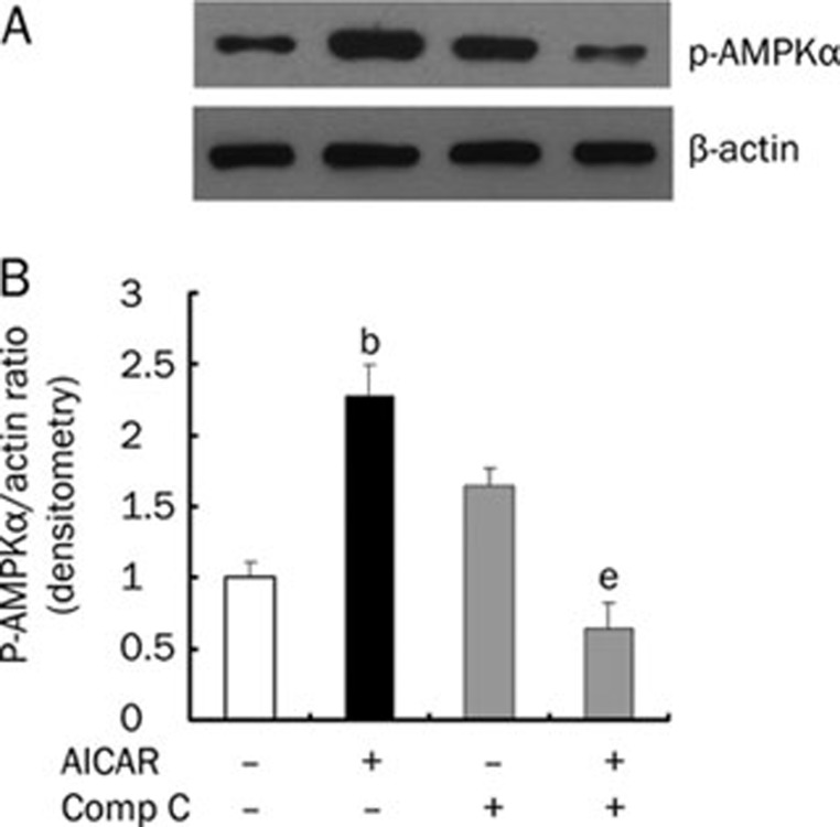
Methods: Rat insulinoma cell line INS-1 was treated with an activator (AICAR) or inhibitor (Compound C) of AMPK as well as inhibitors of PPARs (MK886 to PPARalpha and BADGE to PPARgamma). The mRNA levels of PDX-1, PPARalpha and PPARgamma were measured using real-time RT-PCR, and Western blotting was used to detect the protein expression of these factors.

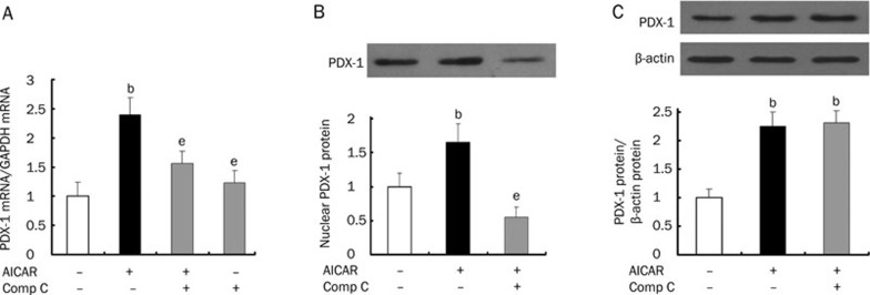
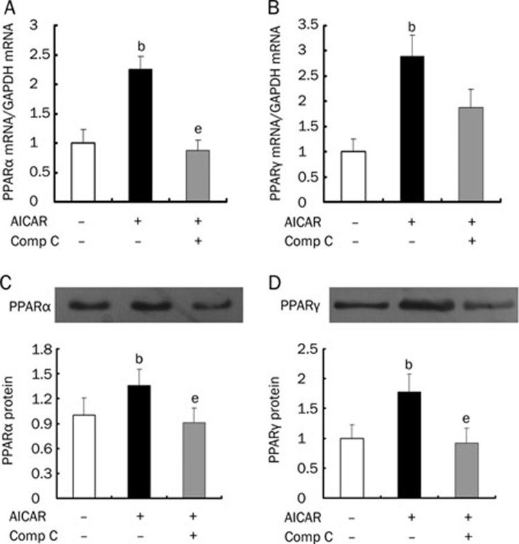
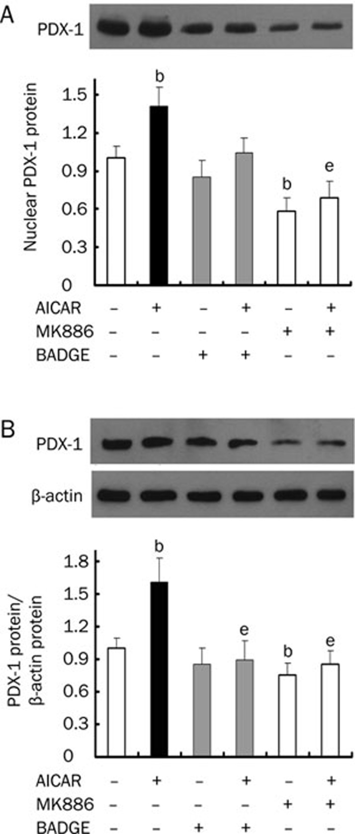
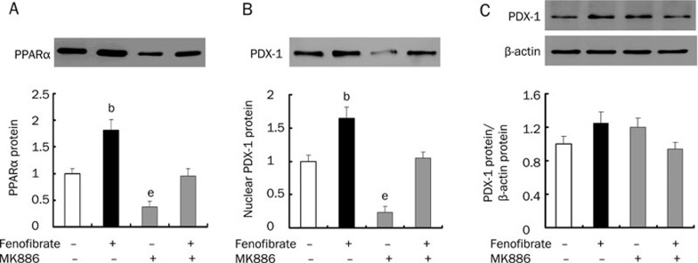
Results: Activation of AMPK by AICAR induced significantly increased the expression of PDX-1, and this increase was abrogated when AMPK was inactivated by Compound C. Similarly, the expression of PPARalpha and PPARgamma was also increased by AICAR or decreased by Compound C. However AMPK activation did not increase nuclear PDX-1 protein levels when PPARalpha was inhibited. In contrast, AMPK activation still up-regulated PDX-1 protein levels during PPARgamma inhibition. Additionally, PPARalpha activation induced by fenofibrate significantly enhanced nuclear PDX-1 protein expression.

Conclusion: AMPK regulates the expression of PDX-1 at both the transcriptional and protein levels, and PPARalpha may be acutely involved in the regulation of INS-1 cells.


Figures

Figure 1
Figure 1
AICAR stimulates the activity of AMPKα. INS-1 cells treated with or without 0.5 mmol/L AICAR in the presence or absence of 10 μmol/L Compound C (Comp C) for 8 h were washed with phosphate-buffered saline and were lysed. For each sample, 60 μg protein was used for Western blotting. (A) A representative blot is shown. (B) Densitometric results from six independent Western blotting experiments are shown. Data were calculated using β-actin as an internal standard and were normalized by setting the control as 1 (mean±SD). bP<0.05 compared to the untreated control group; eP<0.05 compared to the group treated with AICAR alone.
Figure 2
Figure 2
AMPK activation increases the expression of PDX-1. INS-1 cells were treated with or without 0.5 mmol/l AICAR in the presence or absence of 10 μmol/L Compound C (Comp C) for 8 h, and were then harvested to monitor mRNA levels of PDX-1 (A) by real-time RT-PCR. Data were calculated using GAPDH as an internal standard and were normalized by setting the control as 1 (mean±SD, n=6). PDX-1 protein from nuclear (B) and cytoplasmic (C) fractions were detected by Western blotting. Densitometric results from six independent Western blotting experiments are shown. Data were calculated using β-actin as an internal standard and were normalized by setting the control as 1 (mean±SD, n=6). bP<0.05 compared to the untreated control group; eP<0.05 compared to the group treated with AICAR alone.
Figure 3
Figure 3
AICAR up-regulates PPARα and PPARγ expression. INS-1 cells were treated with or without 0.5 mmol/L AICAR, in the presence or absence of 10 μmol/L Compound C (Comp C) for 8 h. They were then harvested to monitor mRNA levels of PPARα (A) and PPARγ (B) by real-time RT-PCR. Results from six independent experiments are shown. Data were calculated using GAPDH as an internal standard and were normalized by setting the control as 1 (mean±SD). Nuclear protein fractions were prepared to detect PPARα (C) and PPARγ (D) by immunoprecipitation. Densitometric results from six independent Western blotting experiments are shown. Data were normalized by setting the control as 1 (mean±SD). bP<0.05 compared to the untreated control group; eP<0.05 compared to the group treated with AICAR alone.
Figure 4
Figure 4
Effects of MK886 and BADGE on the expression of PDX-1. INS-1 cells were pre-incubated with 5 μmol/L MK 886 or 50 μmol/L BADGE for 16 h and in the presence or absence of 0.5 mmol/L AICAR for 8 h. PDX-1 protein from nuclear (A) and cytoplasmic (B) fractions was detected by Western blotting. Densitometric results from six independent Western blotting experiments are shown. Data were calculated using β-actin as an internal standard and were normalized by setting the control as 1 (mean±SD, n=6). bP<0.05 compared to the untreated control group; eP<0.05 compared to the group treated with AICAR alone.
Figure 5
Figure 5
Effects of fenofibrate on the expression of PDX-1 and PPARα. INS-1 cells were treated with or without 5 μmol/L fenofibrate in the presence or absence of 5 μmol/L MK886 for 8 h. Nuclear PPARα protein (A) was detected by immunoprecipitation, and PDX-1 protein from nuclear (B) and cytoplasmic (C) fractions was detected by Western blotting. Densitometric results from six independent Western blotting experiments are shown. bP<0.05 compared to the untreated control group; eP<0.05 compared to the group treated with fenofibrate alone.

Similar articles

Cited by

References

    1. Merrill GF, Kurth EJ, Hardie DG, Winder WW. AICA riboside increases AMP-activated protein kinase, fatty acid oxidation, and glucose uptake in rat muscle. Am J Physiol. 1997;273:E1107–12. - PubMed
    1. Kurth-Kraczek EJ, Hirshman MF, Goodyear LJ, Winder WW. 5′AMP-activated protein kinase activation causes GLUT4 translocation in skeletal muscle. Diabetes. 1999;48:1667–71. - PubMed
    1. Ojuka EO, Jones TE, Nolte LA, Chen M, Wamhoff BR, Sturek M, et al. Regulation of GLUT4 biogenesis in muscle: evidence for involvement of AMPK and Ca2+ Am J Physiol Endocrinol Metab. 2002;282:E1008–13. - PubMed
    1. Leff T. AMP-activated protein kinase regulates gene expression by direct phosphorylation of nuclear proteins. Biochem Soc Trans. 2003;31:224–7. - PubMed
    1. Saleh MC, Fatehi-Hassanabad Z, Wang R, Nino-Fong R, Wadowska DW, Wright GM, et al. Mutated ATP synthase induces oxidative stress and impaired insulin secretion in beta-cells of female BHE/cdb rats. Diabetes Metab Res Rev. 2008;24:392–403. - PubMed

Publication types

MeSH terms