. 2010 Jul 13;5(7):e11513.
doi: 10.1371/journal.pone.0011513.

All-trans retinoic acid directs urothelial specification of murine embryonic stem cells via GATA4/6 signaling mechanisms

Affiliations

All-trans retinoic acid directs urothelial specification of murine embryonic stem cells via GATA4/6 signaling mechanisms

Joshua R Mauney et al. PLoS One. .

Abstract

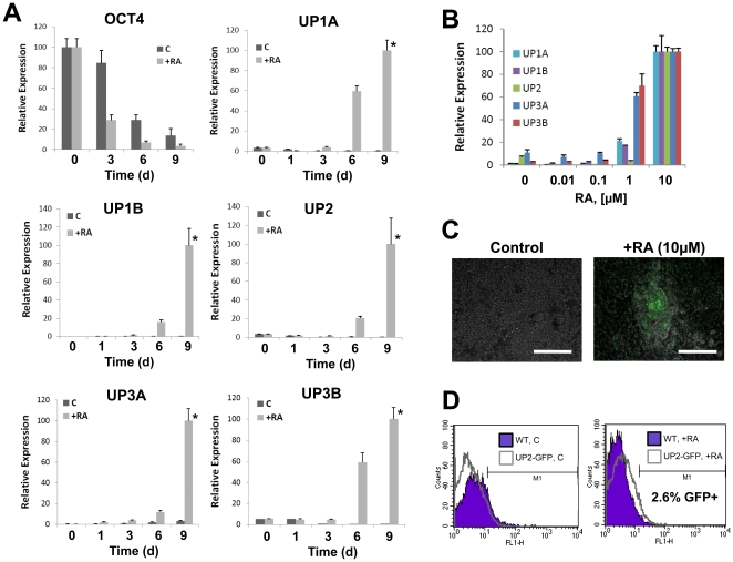
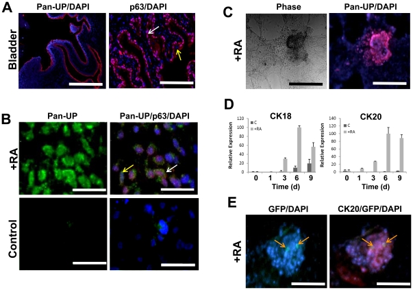
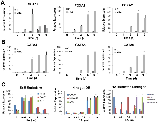
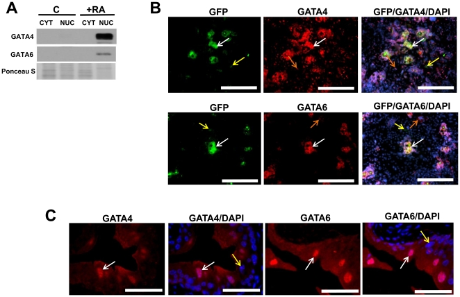
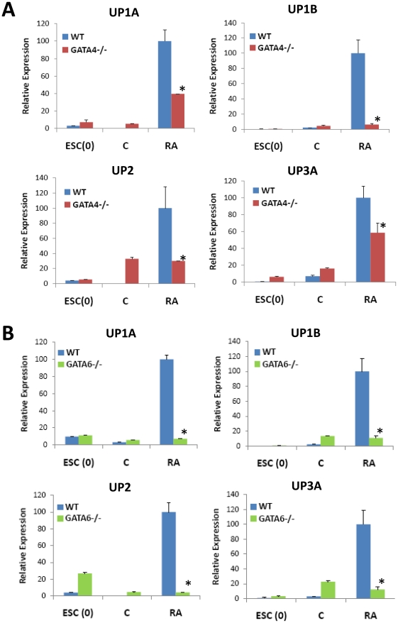
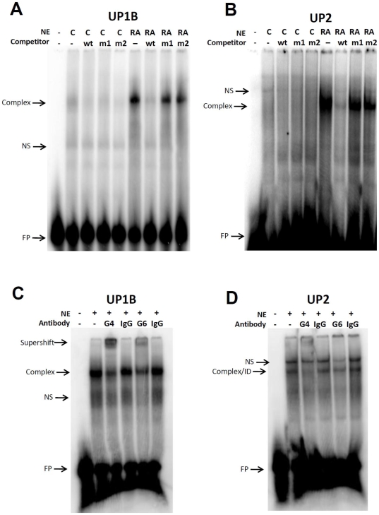
The urinary bladder and associated tract are lined by the urothelium, a transitional epithelium that acts as a specialized permeability barrier that protects the underlying tissue from urine via expression of a highly specific group of proteins known as the uroplakins (UP). To date, our understanding of the developmental processes responsible for urothelial differentiation has been hampered due to the lack of suitable models. In this study, we describe a novel in vitro cell culture system for derivation of urothelial cells from murine embryonic stem cells (ESCs) following cultivation on collagen matrices in the presence all trans retinoic acid (RA). Upon stimulation with micromolar concentrations of RA, ESCs significantly downregulated the pluripotency factor OCT-4 but markedly upregulated UP1A, UP1B, UP2, UP3A, and UP3B mRNA levels in comparison to naïve ESCs and spontaneously differentiating controls. Pan-UP protein expression was associated with both p63- and cytokeratin 20-positive cells in discrete aggregating populations of ESCs following 9 and 14 days of RA stimulation. Analysis of endodermal transcription factors such as GATA4 and GATA6 revealed significant upregulation and nuclear enrichment in RA-treated UP2-GFP+ populations. GATA4-/- and GATA6-/- transgenic ESC lines revealed substantial attenuation of RA-mediated UP expression in comparison to wild type controls. In addition, EMSA analysis revealed that RA treatment induced formation of transcriptional complexes containing GATA4/6 on both UP1B and UP2 promoter fragments containing putative GATA factor binding sites. Collectively, these data suggest that RA mediates ESC specification toward a urothelial lineage via GATA4/6-dependent processes.


Conflict of interest statement

Competing Interests: The authors have declared that no competing interests exist.

Figures

Figure 1
Figure 1. RA stimulation of ESCs induces UP expression in a time and concentration-dependent manner.
[A] Real time RT-PCR analysis of UP and OCT-4 expression by ESCs cultured on collagen matrices in the presence (+RA) or absence (C) of 10 µM RA for up to 9 d. (*) = p<0.05, in comparison to spontaneously differentiating controls and undifferentiated ESCs. [B] Effect of RA concentration on UP expression following 9 d. For [A–B], levels normalized to GAPDH expression. Mean ± SD per data point. [C] Photomicrographs of merged phase and GFP expression in spontaneously differentiating controls and RA-stimulated UP2-GFP ESC line following 14 d of cultivation. Scale bar = 80 µm. [D] Flow cytometry histograms comparing the extent of GFP expression in untransfected (WT) and UP2-GFP ESC lines either as spontaneously differentiating controls (left panel) or RA-treated cultures following 14 d (right panel). Extent of fluorescence intensity per amount of population events is displayed with shaded plots representing WT cultures and open plots representing UP2-GFP ESC line.
Figure 2
Figure 2. RA enrichment of UP+ populations coincides with markers of stratified urothelium.
[A] Photomicrographs of adult murine bladder (C57BL/6) showing native urothelium organization and architecture. Left panel: immunofluorescence of pan-UP expression (red, Cy3) localized in urothelium. Scale bar = 500 µm. Right panel: immunofluorescence of nuclear p63 expression (red, Cy3) localized to the basal and intermediate urothelial layers (denoted with white arrow). Absence of nuclear p63 staining was noted in superficial urothelial cells (denoted with yellow arrow). Scale bar = 100 µm. For both panels, images were merged with DAPI nuclear counterstain (blue). [B] Photomicrographs of spontaneously differentiating control and RA-treated ESCs following 9 d of cultivation showing acquisition of epithelial morphology and co-localization of UP (green, FITC) and nuclear p63 expression (red, Cy3). Pan-UP+ populations merged with DAPI were observed with (denoted with white arrow) and without (denoted with yellow arrow) p63+ nuclear staining. Scale bar = 30 µm. [C] Photomicrographs of phase and immunofluorescence fields of RA-treated ESCs following 14 d of cultivation. Samples demonstrated 3-D cell aggregates coupled with pan-UP expression (red, Cy3). DAPI nuclear counterstain (blue). Scale bar = 100 µm. [D] Real time RT-PCR analysis of CK18 and CK20 expression in cultures described in [B]. Levels normalized to GAPDH expression. Mean ± SD per data point. [E] Photomicrographs of immunofluorescence fields of RA-treated UP2-GFP ESCs following 14 d of cultivation demonstrated GFP expression colocalized with CK20 expression (red, Cy3). Cells exhibiting co-staining of GFP and Cy3 are denoted with orange arrows. DAPI nuclear counterstain (blue). Scale bar = 60 µm.
Figure 3
Figure 3. RA induction of UP expression is correlated with markers of hindgut definitive endoderm (DE), in contrast to markers of the extraembryonic endoderm (ExE).
[A, B] Real time RT-PCR analysis of endoderm marker expression by ESCs cultured on collagen matrices in the presence (+RA) or absence (C) of 10 µM RA for up to 9 d. [C] Effect of RA concentration on ESC differentiation markers of ExE or hindgut DE, as well as various RA-responsive lineages following 9 d of stimulation. For all panels, levels were normalized to GAPDH expression. N = 3–4 per data point.
Figure 4
Figure 4. Nuclear GATA4 and GATA6 expression is associated with UP expression both in vitro and in vivo.
[A] Immunoblot analysis of nuclear (NUC) and cytoplasmic (CYT) protein fractions demonstrating enrichment of GATA4 and GATA6 in nuclear extracts of wild type (WT) RA-treated ESCs following 9 d of cultivation. The degree of Ponceau S staining of membranes after transfer was used to indicate uniform sample loading. [B] Photomicrographs of RA-treated UP2-GFP+ cells co-stained for GFP (green, FITC) and nuclear GATA4/6 (red, Cy3) following 14 d of culture. Observed populations included: GFP/GATA4/6+ cells (denoted by white arrows), cells positive for GATA4/6 expression alone (denoted by orange arrows), and GFP+, but GATA4/6 negative cell types (denoted by yellow arrows). Scale bar = 500 µm. [C] Photomicrographs of adult murine (C57BL/6) bladder urothelium showing nuclear localization of GATA4/6 in superficial cells (denoted by white arrows), while exclusion in the basal and intermediate cell layers (denoted by yellow arrows). Scale bar = 40 µm. For both [A, B], images were merged with DAPI nuclear counterstain (blue).
Figure 5
Figure 5. GATA4 and GATA6 are crucial signaling molecules in RA-mediated upregulation of UP expression in ESCs.
Real time RT-PCR analysis of uroplakin expression in WT, GATA4−/− [A] or GATA6−/− [B] ESCs cultured in the presence (+RA) or absence (C) of 10 µM RA for up to 9 d. ESC(0)  =  undifferentiated ESCs. Levels normalized to GAPDH expression. Mean ± SD per data point. N = 3–4 per data point. (*) = p<0.05, in comparison to levels observed with RA-treated WT cultures.
Figure 6
Figure 6. Murine UP1B and UP2 promoters contain GATA-DNA binding sites which recruit complexes containing GATA4/6 in response to RA stimulation.
Nuclear extracts from RA-treated ESCs following 9 d show distinct GATA-DNA complexes with 32P-labeled consensus oligos specific for GATA binding to 2 kb UP1B [A, C] or UP2 [B, D] promoter fragments. Complexes were absent in spontaneously differentiating controls (C). Both complexes were inhibited by addition of respective excess unlabeled wild type (wt) but not mutant GATA oligos (m1, m2). [C, D] The presence of GATA4 (G4) and GATA6 (G6) in both the GATA-UP complexes was determined by supershift or immunodepletion (ID) following antibody-specific incubation (G4, G6), but not species-matched isotype control antibodies (IgG). NS represents non specific band. FP denotes excess free probe.

References

    1. Wu XR, Kong XP, Pellicer A, Kreibich G, Sun TT. Uroplakins in urothelial biology, function, and disease. Kidney Int. 2009;75:1153–1165. - PMC - PubMed
    1. Cheng W, Jacobs WB, Zhang JJ, Moro A, Park JH, et al. DeltaNp63 plays an anti-apoptotic role in ventral bladder development. Development. 2006;133:4783–492. - PubMed
    1. Erickson DR, Schwarze SR, Dixon JK, Clark CJ, Hersh MA. Differentiation associated changes in gene expression profiles of interstitial cystitis and control urothelial cells. J Urol. 2008;180:2681–2867. - PubMed
    1. Schlager TA, Grady R, Mills SE, Hendley JO. Bladder epithelium is abnormal in patients with neurogenic bladder due to myelomeningocele. Spinal Cord. 2004;42:163–168. - PubMed
    1. Shanks JH, Iczkowski KA. Divergent differentiation in urothelial carcinoma and other bladder cancer subtypes with selected mimics. Histopathology. 2009;54:885–900. - PubMed

Publication types

MeSH terms