. 2010 Jul 13;5(7):e11565.
doi: 10.1371/journal.pone.0011565.

Efficacy and safety of inhaled carbon monoxide during pulmonary inflammation in mice

Affiliations

Efficacy and safety of inhaled carbon monoxide during pulmonary inflammation in mice

Michael R Wilson et al. PLoS One. .

Abstract

Background: Pulmonary inflammation is a major contributor to morbidity in a variety of respiratory disorders, but treatment options are limited. Here we investigate the efficacy, safety and mechanism of action of low dose inhaled carbon monoxide (CO) using a mouse model of lipopolysaccharide (LPS)-induced pulmonary inflammation.

Methodology: Mice were exposed to 0-500 ppm inhaled CO for periods of up to 24 hours prior to and following intratracheal instillation of 10 ng LPS. Animals were sacrificed and assessed for intraalveolar neutrophil influx and cytokine levels, flow cytometric determination of neutrophil number and activation in blood, lung and lavage fluid samples, or neutrophil mobilisation from bone marrow.

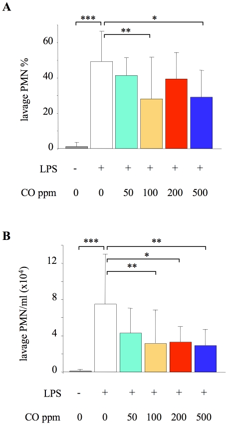
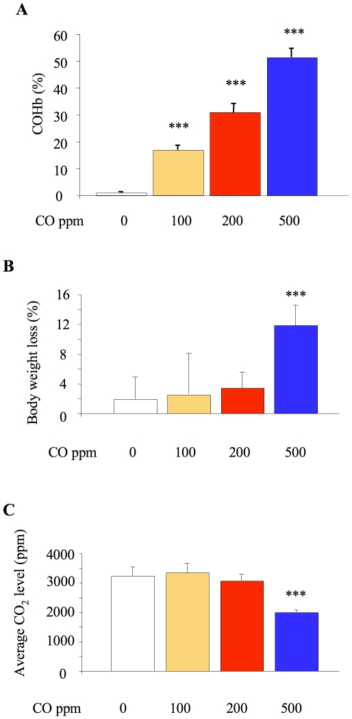
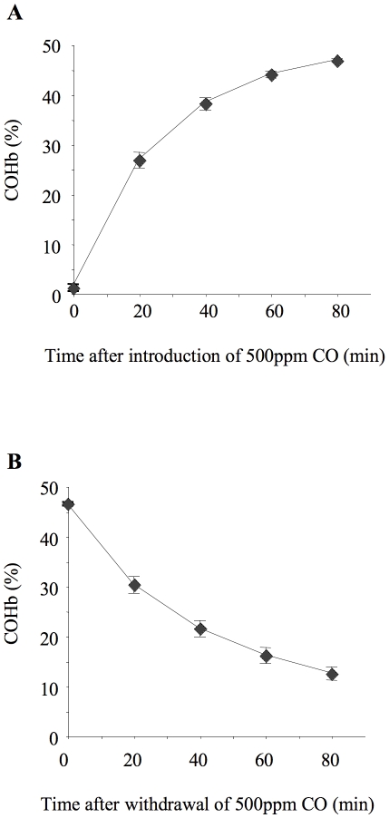
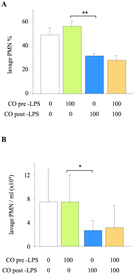
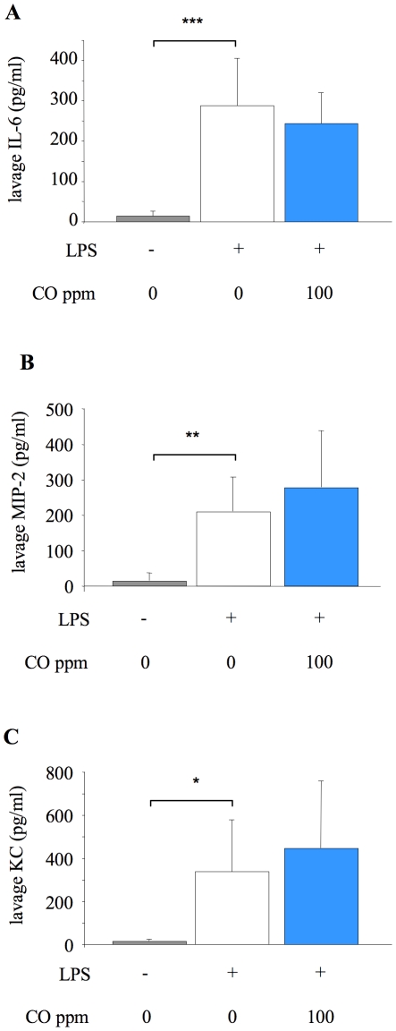
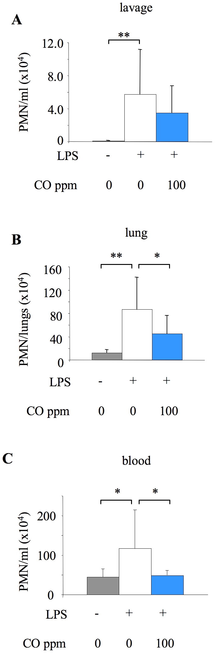
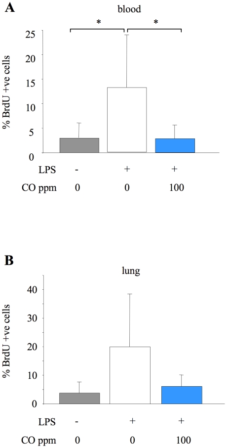
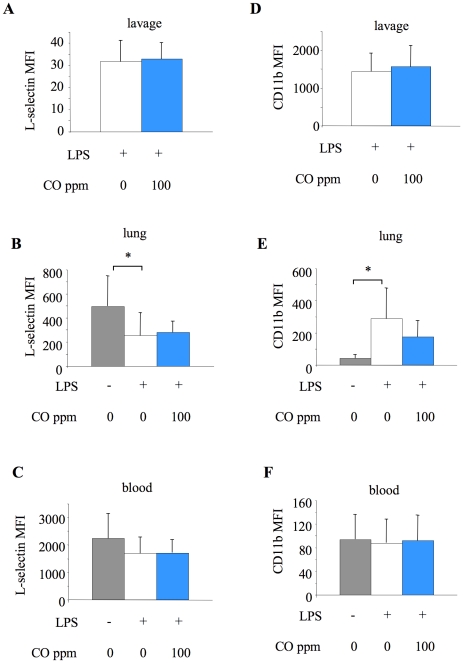
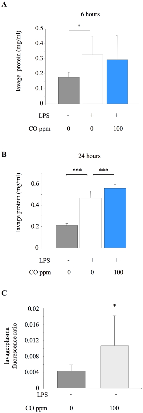
Principal findings: When administered for 24 hours both before and after LPS, inhaled CO of 100 ppm or more reduced intraalveolar neutrophil infiltration by 40-50%, although doses above 100 ppm were associated with either high carboxyhemoglobin, weight loss or reduced physical activity. This anti-inflammatory effect of CO did not require pre-exposure before induction of injury. 100 ppm CO exposure attenuated neutrophil sequestration within the pulmonary vasculature as well as LPS-induced neutrophilia at 6 hours after LPS, likely due to abrogation of neutrophil mobilisation from bone marrow. In contrast to such apparently beneficial effects, 100 ppm inhaled CO induced an increase in pulmonary barrier permeability as determined by lavage fluid protein content and translocation of labelled albumin from blood to the alveolar space.

Conclusions: Overall, these data confirm some protective role for inhaled CO during pulmonary inflammation, although this required a dose that produced carboxyhemoglobin values close to potentially toxic levels for humans, and increased lung permeability.


Conflict of interest statement

Competing Interests: This study was partly supported by a grant from Carburos Metalicos, who have a potential interest in the clinical application of inhaled CO, including salary costs for one of the authors (MG). Carburos Metalicos played no role in study design, data collection and analysis, or writing the manuscript. Authors declare that they have no other competing interests. This does not alter the authors' adherence to all the PLoS ONE policies on sharing data and materials.

Figures

Figure 1
Figure 1. Alveolar neutrophil recruitment 24 hours after LPS.
Impact of carbon monoxide (CO) exposure on neutrophil (PMN) percentage (A) and number/ml (B) in lung lavage fluid of untreated animals (no LPS or CO), or mice treated with 10 ng intratracheal LPS. LPS-challenged mice were exposed to either 0 (air), 50, 100, 200, or 500 ppm CO for 24 hours both before and after LPS. *p<0.05, **p<0.01 ***p<0.001 vs LPS +0 ppm CO; n = 19 for LPS +0 ppm CO, and 8–12 for all other groups (numbers are higher in the LPS+0 ppm CO group because, as our primary control, we ran 1–2 of these animals alongside the experiments for each of the other groups).
Figure 2
Figure 2. Indicators of side-effects with low dose inhaled carbon monoxide.
A. Carboxyhemoglobin (COHb) level in blood of animals exposed to carbon monoxide (CO) for 24 hours both before and after lipopolysaccharide (LPS) instillation. Only COHb data from the first mouse removed from the chamber are shown to minimise the confounding effects of dropping the CO concentration upon opening the chamber. ***p<0.001 vs 0 ppm CO, n = 4–5/group. B. Percentage weight loss in the 24 hours following LPS instillation, in mice exposed to 0, 100, 200 or 500 ppm. ***p<0.001 vs 0 ppm CO, n = 8–12/group. C. CO2 level in chamber. CO2 levels were recorded every 30 minutes: data represent average level in the 24 hour period prior to LPS instillation (to avoid potential confounding effects of anesthetic/LPS). ***p<0.001 vs 0 ppm CO, n = 3–5 experiments, with 4 mice in the chamber each experiment.
Figure 3
Figure 3. Carboxyhemoglobin association and dissociation kinetics.
Time course for association (A) and dissociation (B) of blood carboxyhemoglobin (COHb) in ventilated, instrumented mice. For association kinetics, mice were ventilated from time 0 with 500 ppm carbon monoxide (CO) and arterial blood samples taken every 20 minutes. For dissociation kinetics a separate set of mice were ventilated for 80 minutes with 500 ppm CO, then at time 0 inspired gas was switched to 0 ppm CO and samples were taken every 20 minutes thereafter. n = 4/time point.
Figure 4
Figure 4. Impact of CO exposure either pre- or post- LPS challenge on alveolar neutrophil recruitment.
Neutrophil (PMN) % (A) and number/ml (B) in lung lavage fluid of mice exposed to 100 ppm carbon monoxide (CO) for 24 hours either before or after lipopolysaccharide (LPS) instillation. *p<0.05, **p<0.01 vs 100 ppm CO pre-LPS; n = 8/group. For comparison, data from Figure 1 of the animals exposed either to 0 ppm or 100 ppm CO for 24 hours both pre- and post-LPS are shown (but not included in statistical analysis).
Figure 5
Figure 5. Lavage fluid cytokine concentrations 6 hours after LPS challenge.
Concentration of cytokines IL-6 (A), MIP-2 (B), and KC (C) in lung lavage fluid of untreated mice (no LPS or CO), and mice exposed to 0 or 100 ppm carbon monoxide (CO) for 6 hours after LPS instillation. *p<0.05, **p<0.01 ***p<0.001 vs LPS +0 ppm CO; n = 7–8/group for IL-6 and MIP-2; n = 14–15/group for KC.
Figure 6
Figure 6. Tissue neutrophil numbers 6 hours after LPS, determined by flow cytometry.
Neutrophil (PMN) number in lavage fluid (A), lung tissue (B) and blood (C) from untreated mice (no LPS or CO), or mice exposed to 0 or 100 ppm carbon monoxide (CO) for 6 hours after LPS instillation. Single cell suspensions were prepared from excised lungs of mice by mechanical disruption. Lavage, lung and blood cell samples were stained with fluorochrome-conjugated antibodies against cell-surface markers (CD11b, F4/80, Gr-1, L-selectin) and analysed by flow cytometry. Microsphere counting beads were added to enable cell quantification. Neutrophils were identified based on forward/side-scatter properties and F4/80 and Gr-1 expression. *p<0.05, **p<0.01 vs LPS +0 ppm CO; n = 9–10/group.
Figure 7
Figure 7. Neutrophil mobilisation.
Percentage of newly released BrdU containing neutrophils in blood (A) and lung tissue (B) from untreated mice (no LPS or CO), or mice exposed to 0 or 100 ppm carbon monoxide (CO) for 6 hours after LPS instillation. Neutrophils were identified as described previously, and BrdU incorporated into DNA was detected by flow cytometry using an APC-labelled anti-BrdU antibody. Data are expressed as percentage of neutrophils within tissue positive for BrdU staining. *p<0.05 vs LPS +0 ppm CO; n = 6–7/group.
Figure 8
Figure 8. Neutrophil adhesion molecule expression, determined by flow cytometry.
Surface expression of L-selectin (A–C) and CD11b (D–F) on neutrophils from lavage (A,D), lung tissue (B,E) and blood (C,F) from untreated animals (no LPS or CO), or mice exposed to 0 or 100 ppm carbon monoxide (CO) for 6 hours after LPS instillation. Data are expressed as mean fluorescence intensity (MFI). The data of lavage neutrophils in untreated animals were not included because the numbers of cells recovered were too small to allow for accurate analysis. *p<0.05 vs LPS +0 ppm CO; n = 9–10/group.
Figure 9
Figure 9. Impact of CO on pulmonary barrier permeability.
A. Lung lavage fluid total protein concentration from untreated animals (no LPS or CO), or mice exposed to 0 or 100 ppm carbon monoxide (CO) for 6 hours after LPS instillation. *p<0.05 vs LPS +0 ppm CO; n = 8 for untreated animals and 15–16 for LPS treated groups. B. Lung lavage fluid total protein concentration from untreated animals or mice exposed to 0 or 100 ppm CO for 24 hours after LPS instillation. ***p<0.001 vs LPS +0 ppm CO; n = 7–8/group. C. Permeability was also assessed in both untreated mice (no LPS or CO) and animals receiving 100 ppm CO alone for 6 hours (no LPS) by determining translocation of a fluorescence-labelled albumin from plasma to alveolar space over a 1 hour period. Data are expressed as a ratio of fluorescence between lavage fluid and plasma. *p<0.05 vs untreated group; n = 6–7.

Similar articles

Cited by

References

    1. Ryter SW, Alam J, Choi AM. Heme oxygenase-1/carbon monoxide: from basic science to therapeutic applications. Physiol Rev. 2006;86:583–650. - PubMed
    1. Zhou Z, Song R, Fattman CL, Greenhill S, Alber S, et al. Carbon monoxide suppresses bleomycin-induced lung fibrosis. Am J Pathol. 2005;166:27–37. - PMC - PubMed
    1. Chapman JT, Otterbein LE, Elias JA, Choi AM. Carbon monoxide attenuates aeroallergen-induced inflammation in mice. Am J Physiol Lung Cell Mol Physiol. 2001;281:L209–216. - PubMed
    1. Dolinay T, Szilasi M, Liu M, Choi AM. Inhaled carbon monoxide confers antiinflammatory effects against ventilator-induced lung injury. Am J Respir Crit Care Med. 2004;170:613–620. - PubMed
    1. Kohmoto J, Nakao A, Stolz DB, Kaizu T, Tsung A, et al. Carbon monoxide protects rat lung transplants from ischemia-reperfusion injury via a mechanism involving p38 MAPK pathway. Am J Transplant. 2007;7:2279–2290. - PubMed

Publication types