. 2010 Jul 14;5(7):e11576.
doi: 10.1371/journal.pone.0011576.

Activation of hypoxia inducible factor 1 is a general phenomenon in infections with human pathogens

Affiliations

Activation of hypoxia inducible factor 1 is a general phenomenon in infections with human pathogens

Nadine Werth et al. PLoS One. .

Abstract

Background: Hypoxia inducible factor (HIF)-1 is the key transcriptional factor involved in the adaptation process of cells and organisms to hypoxia. Recent findings suggest that HIF-1 plays also a crucial role in inflammatory and infectious diseases.

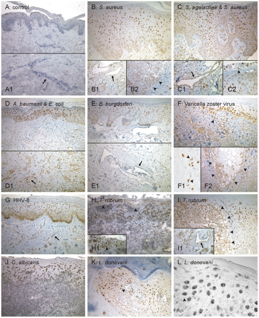
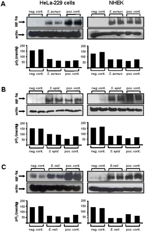
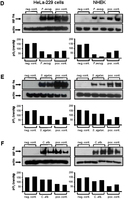
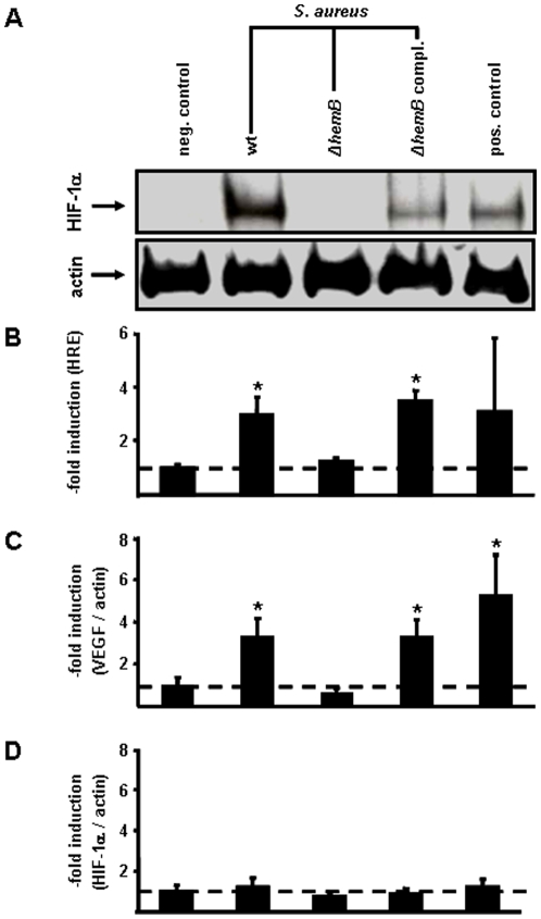
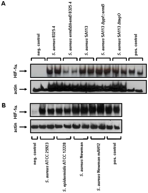
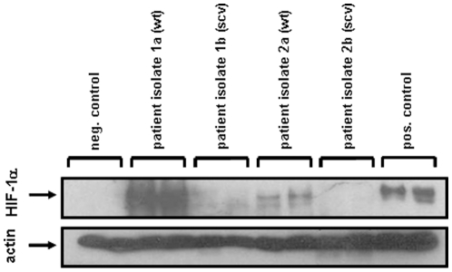
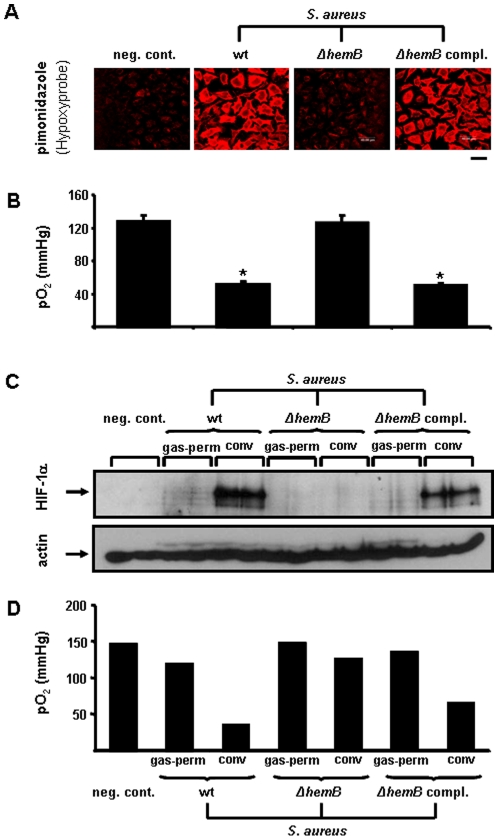
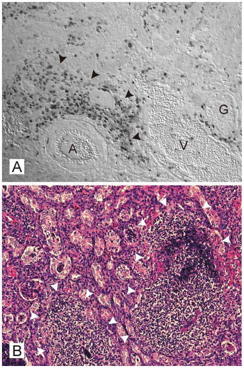
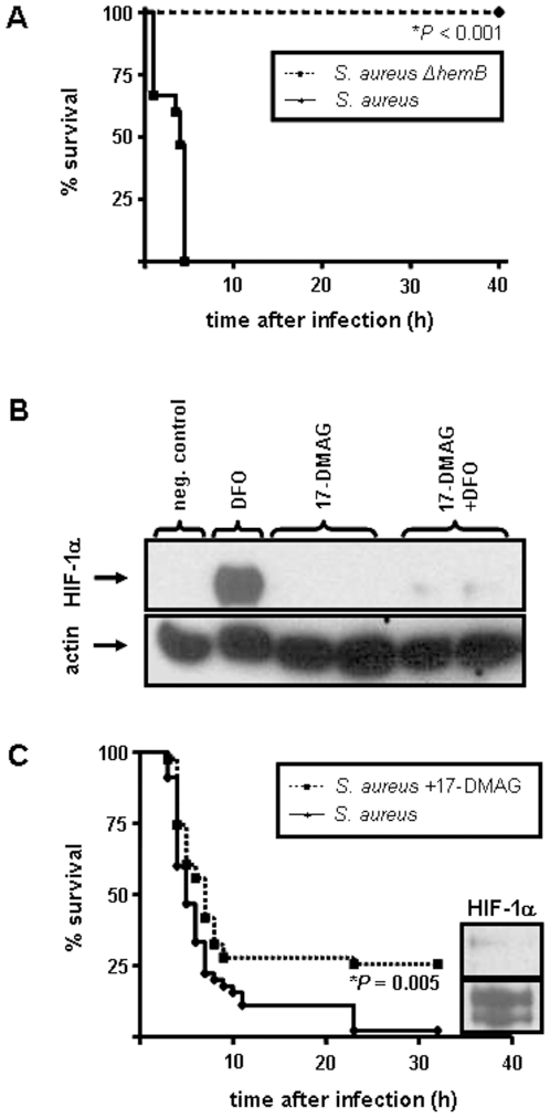
Methodology/principal findings: Using patient skin biopsies, cell culture and murine infection models, HIF-1 activation was determined by immunohistochemistry, immunoblotting and reporter gene assays and was linked to cellular oxygen consumption. The course of a S. aureus peritonitis was determined upon pharmacological HIF-1 inhibition. Activation of HIF-1 was detectable (i) in all ex vivo in biopsies of patients suffering from skin infections, (ii) in vitro using cell culture infection models and (iii) in vivo using murine intravenous and peritoneal S. aureus infection models. HIF-1 activation by human pathogens was induced by oxygen-dependent mechanisms. Small colony variants (SCVs) of S. aureus known to cause chronic infections did not result in cellular hypoxia nor in HIF-1 activation. Pharmaceutical inhibition of HIF-1 activation resulted in increased survival rates of mice suffering from a S. aureus peritonitis.

Conclusions/significance: Activation of HIF-1 is a general phenomenon in infections with human pathogenic bacteria, viruses, fungi and protozoa. HIF-1-regulated pathways might be an attractive target to modulate the course of life-threatening infections.


Conflict of interest statement

Competing Interests: The authors have declared that no competing interests exist.

Figures

Figure 1
Figure 1. Detection of HIF-1 activation in human skin biopsies of patients suffering from cutaneous infections by immunohistochemistry.
(A) In uninfected skin, rare and faint nuclear HIF-1α staining occurred in the epidermis whereas the dermis was negative for HIF-1α (A1). (B to L) In all cutaneous infections, a strong nuclear HIF-1α signal was detected in keratinocytes, predominantly of the spinal cell layer (examplified in L). Moreover, endothelial cells of dermal capillaries frequently stained positive (arrows in B1, C1, D1, E1, G, I1). Arrowheads: (B2) border zone of the positive abscess; (C2): positive neutrophils; (F): positive dermal lympho-histiocytic infiltrates; (F1): large positive nuclei of a multi-nuclear giant cell; (F2): skin lesion surrounded by a HIF-1 positive inflammatory infiltrate; (H): positive sub-corneal neutrophils (higher power in H1); (I): positive intra-epidermal infiltrate; (K): intradermal nodules containing leishmaniae. (A) Control: uninfected sample; (B–E): bioptic samples of patients suffering from infections with humanpathogenic bacteria [(B) S. aureus, (C) S. agalactiae and S. aureus, (D) A. baumanii and E. coli and (E) B. burgdorferi], (F, G) with humanpathogenic Herpes-viruses [(F) Varicella zoster virus (VZV) and (G) Human Herpes Virus-8 (HHV 8)], (H–J) with humanpathogenic fungi [(H+I) T. rubrum and (J) C. albicans] or (K, L) with the protozoic pathogen L. donovani. Magnifications: all 250×, except for A1, B1, B2, C1, D1, E1, F2, I1 (all 400×), and C2, F1, H1, L (all 1,000×).
Figure 2
Figure 2. Oxygen-dependent HIF-1 activation in HeLa-229 cells and NHEKs (Normal Human Epidermal Keratinocytes) by bacterial and fungal pathogens.
Four to six hours upon infection, HIF-1 activation was determined by Western blotting (loading control: actin). pO2 levels were quantified in the medium of control and infected cells (A: S. aureus ATCC 33592, MOI 20, infection time: six hours; B: S. epidermidis ATCC 12228, MOI 20, infection time: six hours; C: E. coli ATCC 25992, MOI 10, infection time: four hours). Negative control: uninfected cells, positive control: hypoxia or DFO (200 µmol/L).
Figure 3
Figure 3. Oxygen-dependent HIF-1 activation in HeLa-229 cells and NHEKs (Normal Human Epidermal Keratinocytes) by bacterial and fungal pathogens.
Four to six hours upon infection, HIF-1 activation was determined by Western blotting (loading control: actin). pO2 levels were quantified in the medium of control and infected cells (D: P. aeruginosa ATCC 27853, MOI 10, infection time: four hours; E: S. agalactiae SK 43, MOI 200, infection time: five hours; F: C. albicans ATCC 90028, MOI 20, infection time: six hours). Negative control: uninfected cells, positive control: hypoxia or DFO (200 µmol/L).
Figure 4
Figure 4. Induction of HIF-1-dependent gene programming in HeLa-229 cells by different S. aureus strains.
Six hours upon infection, HIF-1 activation was analyzed in (A) Western blots (loading control: actin) and by determining (B) the induction of the HIF-1-dependent luciferase activity. HeLa-229 cells were transfected with a 4xHRE-luc reporter construct 24 hours before infection (triplicate means given; for details see Material and Methods). Induction was determined by chemiluminescence. Bacterial induced (C) VEGF and (D) HIF-1α transcription. Total mRNA was prepared six hours upon infection, transcribed into cDNA and quantified by real-time Light-Cycler-PCR (ratio: VEGF/actin or HIF-1α/actin transcripts; triplicate means given). Negative control: uninfected cells; S. aureus wt (8325-4); S. aureus ΔhemB (S. aureus ermBΩhemB 8325-4, SCV); S. aureus ΔhemB, hemB-complemented (S. aureus pCX19ΩhemB 8325-4, complemented SCV mutant) ; positive control: DFO (200 µmol/L). Multiplicity of infection (MOI): 20. * significant difference to control cells (P<0.05).
Figure 5
Figure 5. Induction of HIF-1 activation in HeLa-229 cells by different S. aureus strains defective in the expression of cell wall components.
The HIF-1α protein was analyzed in Western blots six hours upon infection (loading control: actin) of HeLa 229 cells with the following bacterial strains: (A) S. aureus 8325-4 (wt), S. aureus ermBΩhemB 8325-4 (SCV), S. aureus SA113 (wt), S. aureus SA113 Δypf::ermB (reduced in lipoteichoic acid synthesis), S. aureus SA113 ΔtagO (defective in producing wall teichoic acid); (B) S. aureus ATCC 25923 (wt), S. epidermidis ATCC 12228 (wt), S. aureus Newman (wt), S. aureus Newman mAH12 (defective in producing the extracellular adherence protein EAP). Negative control: uninfected cells, positive control: DFO (200 µM). Multiplicity of infection (MOI): 20.
Figure 6
Figure 6. Induction of HIF-1 activation in HeLa-229 cells by S. aureus patient isolates (wild-type and the respective small colony variants).
The HIF-1α protein was analyzed in Western blots six hours upon infection (loading control: actin). The following patient isolates were used: S. aureus patient isolate 1 (wt; S. aureus A22223 I), S. aureus patient isolate 2 (clinical SCV; S. aureus A22223 II), S. aureus patient isolate 3 (wt; S. aureus OM 1a), S. aureus patient isolate 4 (clinical SCV; S. aureus OM 1b). Negative control: uninfected cells, positive control: hypoxia. Multiplicity of infection (MOI): 20.
Figure 7
Figure 7. Cellular oxygen consumption and oxygen-dependent HIF-1 activation in HeLa-229 cells infected with different S. aureus strains.
(A) Control and infected HeLa-229 cells were incubated with the hypoxic marker pimonidazole hydrochloride which was visualized by using specific Cy3 labelled antibodies (for details see Materials and Methods). Scale bare: 40 µm. (B) pO2 levels were quantified in the medium of control and infected cells (triplicate means given). (C) Hela-229 cells were incubated in gas permeable (gas-perm) and conventional (conv) culture dishes and HIF 1α protein was analyzed in Western blots (loading control: actin). (D) pO2 levels in the medium of control or S. aureus infected HeLa-229 cells (means of doubles). Negative control: uninfected cells; S. aureus wt (8325-4); S. aureus ΔhemB (S. aureus ermBΩhemB 8325-4, SCV); S. aureus ΔhemB, hemB-complemented (S. aureus pCX19ΩhemB 8325-4, complemented SCV mutant). Multiplicity of infection (MOI): 20. * significant difference to control cells (P<0.05).
Figure 8
Figure 8. HIF-1 activation in kidneys of mice infected with S. aureus.
(A) HIF 1α immunohistochemistry and (B) hematoxilin-eosin staining of renal mouse abscesses induced by intravenous infection with S. aureus seven days upon infection (following sections). HIF 1α positive nuclei are most abundant within the abscesses borders, some interstitial staining also occurs within the renal parenchyma. In addition, renal tubules in the vicinity of the abscess stain also positively (halfed arrow in B). A: arcuate artery, G: glomerulus, V: arcuate vein; arrowheads: margin of abscesses. Magnifications: (A) 250×, (B) 100×.
Figure 9
Figure 9. Role of HIF-1 in infections with S. aureus in a murine peritonitis model.
(A) Survival of NMRI mice after intraperitoneal infection with S. aureus (wt; S. aureus 8325-4; n = 8) or S. aureus ΔhemB (SCV, S. aureus ermΩhemB 8325-4; n = 8). Note the higher susceptibility of mice infected with S. aureus 8325-4 compared to the mice infected with S. aureus ΔhemB. *significant difference: P<0.001. (B) Inhibition of DFO induced HIF-1 activation in HeLa-229 cells by 17-DMAG. Cells were incubated with 17 DMAG (10 µmol/L) for 16 hours following induction of HIF 1 activation by the iron chelator DFO (200 µmol/L) for six hours. HIF 1α protein was analyzed in Western blots (loading control: actin). Negative control: uninfected cells. (C) Survival rate of NMRI mice after intraperitoneal infection with S. aureus (S. aureus 8325-4). One group of mice was treated 24 h and 16 h before infection with the HIF-1 inhibiting compound 17-DMAG (25 mg/g body weight). Note the higher survival rate of 17-DMAG-treated mice (n = 45) compared with control mice (n = 45). *significant difference: P = 0.005 (Kaplan-Meier analysis).

Similar articles

Cited by

References

    1. Shimoda LA, Manalo DJ, Sham JS, Semenza GL, Sylvester JT. Partial HIF-1alpha deficiency impairs pulmonary arterial myocyte electrophysiological responses to hypoxia. Am J Physiol Lung Cell Mol Physiol. 2001;281:202–208. - PubMed
    1. Pugh CW, Ratcliffe PJ. Regulation of angiogenesis by hypoxia: role of the HIF system. Nat Med. 2003;9:677–684. - PubMed
    1. Ivan M, Kondo K, Yang H, Kim W, Valiando J, et al. HIFalpha targeted for VHL-mediated destruction by proline hydroxylation: implications for O2 sensing. Science. 2001;292:464–468. - PubMed
    1. Mazure NM, Brahimi-Horn MC, Berta MA, Benizri E, Bilton RL, et al. HIF-1: master and commander of the hypoxic world. A pharmacological approach to its regulation by siRNAs. Biochem Pharmacol. 2004;68:971–980. - PubMed
    1. Cramer T, Yamanishi Y, Clausen BE, Forster I, Pawlinski R, et al. HIF-1alpha is essential for myeloid cell-mediated inflammation. Cell. 2003;112:645–657. - PMC - PubMed

Publication types

MeSH terms

Substances

LinkOut - more resources