

Pressure driven spinning: A multifaceted approach for preparing nanoscaled functionalized fibers, scaffolds, and membranes with advanced materials
- PMID: 20644675
- PMCID: PMC2905272
- DOI: 10.1063/1.3328092
Pressure driven spinning: A multifaceted approach for preparing nanoscaled functionalized fibers, scaffolds, and membranes with advanced materials
Abstract
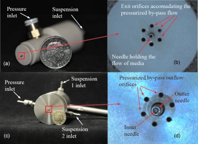
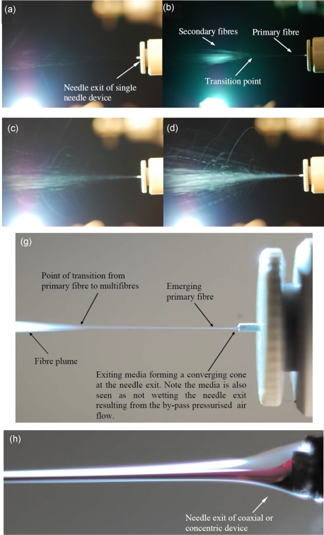
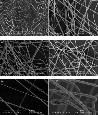
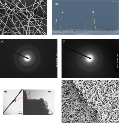
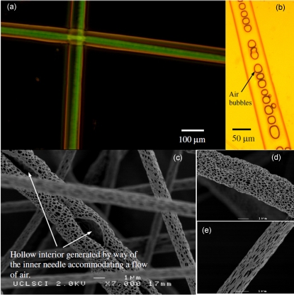
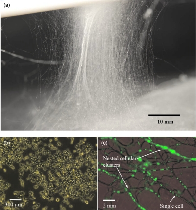
Electrospinning, a flexible jet-based fiber, scaffold, and membrane fabrication approach, has been elucidated as having significance to the heath sciences. Its capabilities have been most impressive as it possesses the ability to spin composite fibers ranging from the nanometer to the micrometer scale. Nonetheless, electrospinning has limitations and hazards, negating its wider exploration, for example, the inability to handle highly conducting suspensions, to its hazardous high voltage. Hence, to date electrospinning has undergone an exhaustive research regime to a point of cliché. Thus, in the work reported herein we unveil a competing technique to electrospinning, which has overcome the above limitations and hazards yet comparable in capabilities. The fiber preparation approach unearthed herein is referred to as "pressure driven spinning (PDS)." The driving mechanism exploited in this fiber spinning process is the pressurized by-pass flow. This mechanism allows the drawing of either micro- or nanosized fibers while processing polymeric suspensions containing a wide range of advanced materials spanning structural, functional, and biological entities. Similar to electrospinning if the collection time of these continuous formed fibers is varied, composite scaffolds and membranes are generated. In keeping with our interests, multicompositional structural entities such as these could have several applications in biology and medicine, for example, ranging from the development of three-dimensional cultures (including disease models) to the development of synthetic tissues and organ structures to advanced approaches for controlled and targeted therapeutics.
Figures
Similar articles
-
A novel direct fibre generation technique for preparing functionalized and compound scaffolds and membranes for applications within the life sciences.Biomed Mater. 2007 Sep;2(3):189-95. doi: 10.1088/1748-6041/2/3/004. Epub 2007 Aug 23. Biomed Mater. 2007. PMID: 18458471
-
Engineering hybrid polymer-protein super-aligned nanofibers via rotary jet spinning.Biomaterials. 2014 Mar;35(10):3188-97. doi: 10.1016/j.biomaterials.2013.12.072. Epub 2014 Jan 20. Biomaterials. 2014. PMID: 24456606
-
Nanostructured degradable macroporous hydrogel scaffolds with controllable internal morphologies via reactive electrospinning.Acta Biomater. 2020 Mar 1;104:135-146. doi: 10.1016/j.actbio.2019.12.038. Epub 2020 Jan 3. Acta Biomater. 2020. PMID: 31904560
-
A review of evolution of electrospun tissue engineering scaffold: From two dimensions to three dimensions.Proc Inst Mech Eng H. 2017 Jul;231(7):597-616. doi: 10.1177/0954411917699021. Epub 2017 Mar 28. Proc Inst Mech Eng H. 2017. PMID: 28347262 Review.
-
A review of developments in electrospinning technology: new opportunities for the design of artificial tissue structures.Int J Artif Organs. 2011 Oct;34(10):986-97. doi: 10.5301/ijao.5000062. Int J Artif Organs. 2011. PMID: 22161282 Review.
Cited by
-
25th anniversary article: Rational design and applications of hydrogels in regenerative medicine.Adv Mater. 2014 Jan 8;26(1):85-123. doi: 10.1002/adma.201303233. Adv Mater. 2014. PMID: 24741694 Free PMC article. Review.
-
Convenient quantification of methanol concentration detection utilizing an integrated microfluidic chip.Biomicrofluidics. 2012 Aug 13;6(3):34111. doi: 10.1063/1.4746246. Print 2012 Sep. Biomicrofluidics. 2012. PMID: 23940501 Free PMC article.
-
Fiber-based tissue engineering: Progress, challenges, and opportunities.Biotechnol Adv. 2013 Sep-Oct;31(5):669-87. doi: 10.1016/j.biotechadv.2012.11.007. Epub 2012 Nov 27. Biotechnol Adv. 2013. PMID: 23195284 Free PMC article. Review.
References
LinkOut - more resources
Full Text Sources
Miscellaneous