. 2010 Jul 14;5(7):e11574.
doi: 10.1371/journal.pone.0011574.

miR-148 regulates Mitf in melanoma cells

Affiliations

miR-148 regulates Mitf in melanoma cells

Benedikta S Haflidadóttir et al. PLoS One. .

Abstract

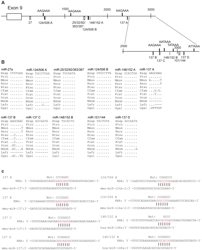
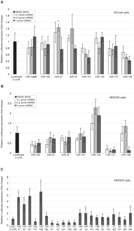
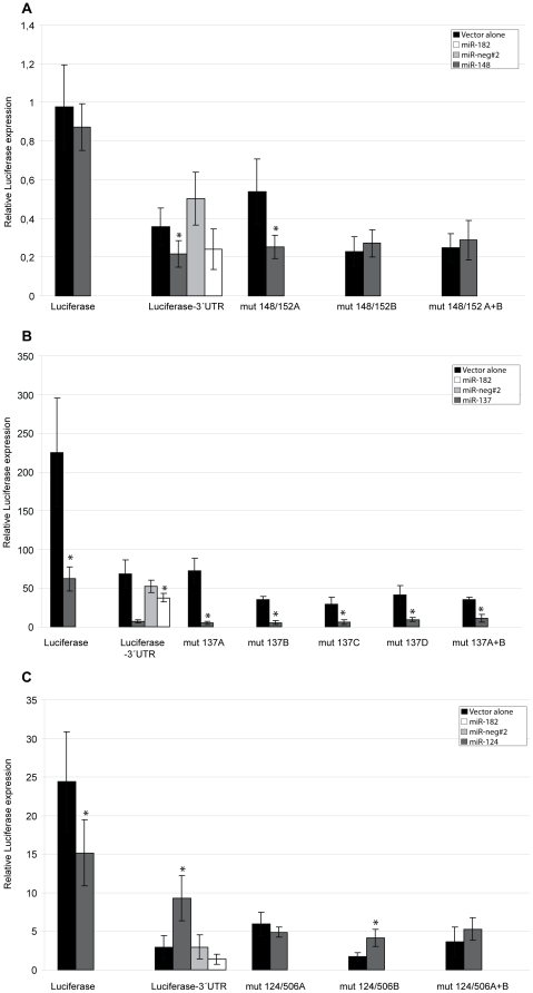
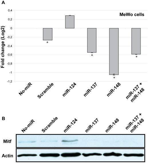
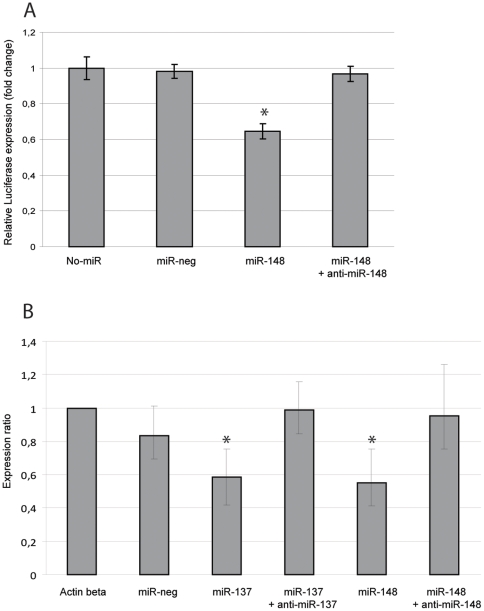
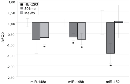
The Microphthalmia associated transcription factor (Mitf) is an important regulator in melanocyte development and has been shown to be involved in melanoma progression. The current model for the role of Mitf in melanoma assumes that the total activity of the protein is tightly regulated in order to secure cell proliferation. Previous research has shown that regulation of Mitf is complex and involves regulation of expression, splicing, protein stability and post-translational modifications. Here we show that microRNAs (miRNAs) are also involved in regulating Mitf in melanoma cells. Sequence analysis revealed conserved binding sites for several miRNAs in the Mitf 3'UTR sequence. Furthermore, miR-148 was shown to affect Mitf mRNA expression in melanoma cells through a conserved binding site in the 3'UTR sequence of mouse and human Mitf. In addition we confirm the previously reported effects of miR-137 on Mitf. Other miRNAs, miR-27a, miR-32 and miR-124 which all have conserved binding sites in the Mitf 3'UTR sequence did not have effects on Mitf. Our data show that miR-148 and miR-137 present an additional level of regulating Mitf expression in melanocytes and melanoma cells. Loss of this regulation, either by mutations or by shortening of the 3'UTR sequence, is therefore a likely factor in melanoma formation and/or progression.


Conflict of interest statement

Competing Interests: The authors have declared that no competing interests exist.

Figures

Figure 1
Figure 1. Schematic presentation of the mouse Mitf -3′UTR sequence.
A. The line indicates the 3′ UTR region of the mouse Mitf gene, including the coding region of exon 9. Potential binding sites for miR-27, miR-124/506, miR-25/32/92/363/367, miR-148/152, miR-137 and miR-101/144 in the mMitf 3′UTR sequence are indicated below the line and potential PAS sites above. Positions are based on the mouse Mitf 3′UTR sequence and are numbered starting at the first nucleotide after the stop codon. Black bars: miR-124/506 binding sites, dark grey bars: miR-137 binding sites, light grey bars: miR-148/152 binding sites, white bars: miR-27, miR-25/32/92/363/367 and miR-101/144. B. The conservation of potential miRNA binding sites in the Mitf 3′UTR sequence. Dots represent a conserved base, short lines represent absence of a sequence at this location. C. Mutations made in the Mitf 3′UTR sequence. The wild type mouse Mitf mRNA sequence is shown with potential binding sites indicated in red. The mature miRNA sequence and potential binding between the miRNA seed region to the 3′UTR sequence are shown and mutated bases are indicated above the mRNA sequence.
Figure 2
Figure 2. Effects of microRNAs on the Mitf-3′UTR-luciferase reporter.
A. Effects of miRNAs on the mouseMitf-3′UTR-luciferase reporter in 501melanoma cells. Three concentrations of miRNAs were tested: 0.1 pmol, 0.5 pmol and 1 pmol. Results in all panels are presented as mean values ±SD. Difference between the mouseMitf- 3′UTR-luciferase vector with and without co-transfected miRNA was considered statistically significant when p<0.05, shown with an asterisk (T-test, 95% confidence level). P-values are presented in Table S1. B. Effects of miRNAs on the mouseMitf- 3′UTR-luciferase vector in HEK293 cells. Three concentrations of miRNAs were tested: 0.1 pmol, 0.5 pmol and 1 pmol. C. Effects of miRNAs when transfected in combination with another miRNA on the reporter. Concentration of each miRNA is 0.5 pmol.
Figure 3
Figure 3. Mutating miRNA binding sites affects the Mitf mRNA.
A. Effects of miR-148 on the mouseMitf-3′UTR-luciferase reporter when the potential binding sites are mutated. Results in all panels are presented as mean values ±SD. Difference between the appropriate vector (labeled on the X-axis) with and without co-transfected miRNA was considered statistically significant when p<0.05, shown with an asterisk (T-test, 95% confidence level). P-values are presented in Table S1. B. Effects of miR-137 on the mouseMitf-3′UTR-luciferase reporter when the potential binding sites are mutated. C. Effects of miR-124 on the mouseMitf-3′UTR-luciferase reporter when the potential binding sites are mutated.
Figure 4
Figure 4. Effects of miRNAs on endogenous MITF.
A. Expression of human MITF mRNA in MeWo cells transfected with miR-124, miR-137, miR-148 or miR-137 and miR-148 combined. The figure shows average expression levels of three replicates for each sample relative to untreated cells (No-miR). Negative control is a scramble miRNA sequence (scramble). Statistically significant difference between untreated cells and treated cells are shown with an asterisk (T-test, 95% confidence level). P-values are: Scramble  = 0.0396; miR-124 = 0.2228; miR-137 = 0.0069; miR-148 = 0.0051; miR-137+148 = 0.0051. B. Western blot analysis on MITF protein levels in MeWo cells, when transfected with miR-124, miR-137, miR-148 or miR-137 and miR-148 combined.
Figure 5
Figure 5. Inhibiting the miRNAs blocks their effect.
A. Co-transfection of anti-miR-148 and miR-148 simultaneously with the Mitf-3′UTR-luciferase vector inhibits the effect of miR-148. Results are presented as mean values ±SD. Expression levels were compared to a sample where no microRNAs were added (No-miR) and were considered statistically significant when p<0.05, shown with an asterisk (T-test, 95% confidence level). The p-values are; miR-neg  = 0.14 miR-148 = 0.007, miR-148+anti-miR-148 = 0.119. B. Transfecting miRNAs and anti-miRNAs blocked the effect of the miRNAs on endogenous human MITF mRNA levels in MeWo cells. P-values when compared to the no-miR sample are: Neg control  = 0.064, miR-137 = 0.006, miR-137 + anti-miR-137 = 0.854, miR-148 = 0.004, miR-148 + anti-miR-148 = 0.632.
Figure 6
Figure 6. Endogenous miRNA expression.
Expression of endogenous miRNAs miR-148a, miR-148b and miR-152 in HEK293, 501mel and MeWo cells. The figure shows average expression levels of three replicates for each sample relative to HEK293 cells. Expression was normalized to miR-16 and miR-103. Statistically significant levels of expression in MeWo and 501mel compared to HEK293 (p<0.05) are shown with an asterisk. P-values are: miR-148a expression in 501mel  = 0.0940 and in MeWo  = 0.0148. miR-148b expression in 501mel  = 0.0023 and in MeWo  = 0.0065. miR-152 expression in 501mel  = 0.0054 and in MeWo =  0.9316.

Similar articles

Cited by

References

    1. Steingrimsson E, Copeland NG, Jenkins NA. Melanocytes and the microphthalmia transcription factor network. Annu Rev Genet. 2004;38:365–411. - PubMed
    1. Nishimura EK, Granter SR, Fisher DE. Mechanisms of hair graying: incomplete melanocyte stem cell maintenance in the niche. Science. 2005;307:720–724. - PubMed
    1. Hodgkinson CA, Moore KJ, Nakayama A, Steingrimsson E, Copeland NG, et al. Mutations at the mouse microphthalmia locus are associated with defects in a gene encoding a novel basic-helix-loop-helix-zipper protein. Cell. 1993;74:395–404. - PubMed
    1. Hughes MJ, Lingrel JB, Krakowsky JM, Anderson KP. A helix-loop-helix transcription factor-like gene is located at the mi locus. J Biol Chem. 1993;268:20687–20690. - PubMed
    1. Hemesath TJ, Steingrimsson E, McGill G, Hansen MJ, Vaught J, et al. microphthalmia, a critical factor in melanocyte development, defines a discrete transcription factor family. Genes Dev. 1994;8:2770–2780. - PubMed

Publication types

MeSH terms

Substances