

The contribution of the Tie2+ lineage to primitive and definitive hematopoietic cells
- PMID: 20645309
- PMCID: PMC2944906
- DOI: 10.1002/dvg.20654
The contribution of the Tie2+ lineage to primitive and definitive hematopoietic cells
Abstract
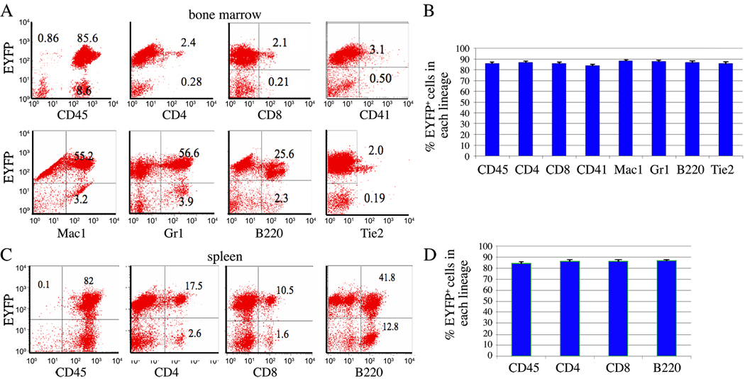
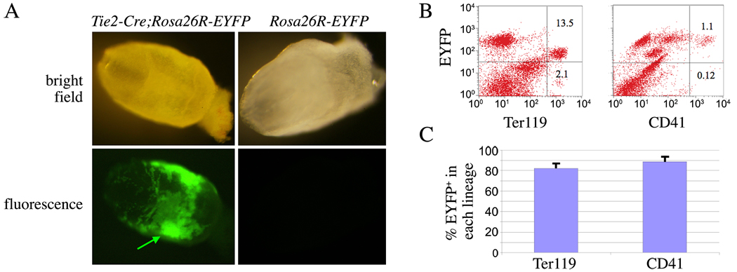
The regulatory elements of the Tie2/Tek promoter are commonly used in mouse models to direct transgene expression to endothelial cells. Tunica intima endothelial kinase 2 (Tie2) is also expressed in hematopoietic cells, although this has not been fully characterized. We determine the lineages of adult hematopoietic cells derived from Tie2-expressing populations using Tie2-Cre;Rosa26R-EYFP mice. In Tie2-Cre;Rosa26R-EYFP mice, analysis of bone marrow cells showed Cre-mediated recombination in 85% of the population. In adult bone marrow and spleen, we analyzed subclasses of early hematopoietic progenitors, T cells, monocytes, granulocytes, and B cells. We found that ∼ 84% of each lineage was EYFP(+), and nearly all cells that come from Tie2-expressing lineages are CD45(+), confirming widespread contribution to definitive hematopoietic cells. In addition, more than 82% of blood cells within the embryonic yolk sac were of Tie2(+) origin. Our findings of high levels of Tie2-Cre recombination in the hematopoietic lineage have implications for the use of the Tie2-Cre mouse as a lineage-restricted driver strain.
© 2010 Wiley-Liss, Inc.
Figures
References
-
- Dumont DJ, Gradwohl G, Fong GH, Puri MC, Gertsenstein M, Auerbach A, Breitman ML. Dominant-negative and targeted null mutations in the endothelial receptor tyrosine kinase, tek, reveal a critical role in vasculogenesis of the embryo. Genes Dev. 1994;8:1897–1909. - PubMed
-
- Eilken HM, Nishikawa S, Schroeder T. Continuous single-cell imaging of blood generation from haemogenic endothelium. Nature. 2009;457:896–900. - PubMed
-
- Ema M, Yokomizo T, Wakamatsu A, Terunuma T, Yamamoto M, Takahashi S. Primitive erythropoiesis from mesodermal precursors expressing VE-cadherin, PECAM-1, Tie2, endoglin, and CD34 in the mouse embryo. Blood. 2006;108:4018–4024. - PubMed
-
- Forde A, Constien R, Grone HJ, Hammerling G, Arnold B. Temporal Cre-mediated recombination exclusively in endothelial cells using Tie2 regulatory elements. Genesis. 2002;33:191–197. - PubMed
Publication types
MeSH terms
Substances
Grants and funding
LinkOut - more resources
Full Text Sources
Medical
Molecular Biology Databases
Research Materials
Miscellaneous
