

Altered intrinsic functional connectivity of anterior and posterior insula regions in high-functioning participants with autism spectrum disorder
- PMID: 20645311
- PMCID: PMC6870194
- DOI: 10.1002/hbm.21085
Altered intrinsic functional connectivity of anterior and posterior insula regions in high-functioning participants with autism spectrum disorder
Abstract
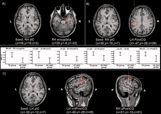
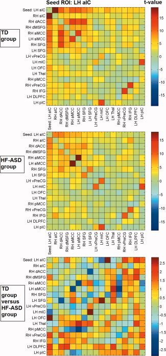
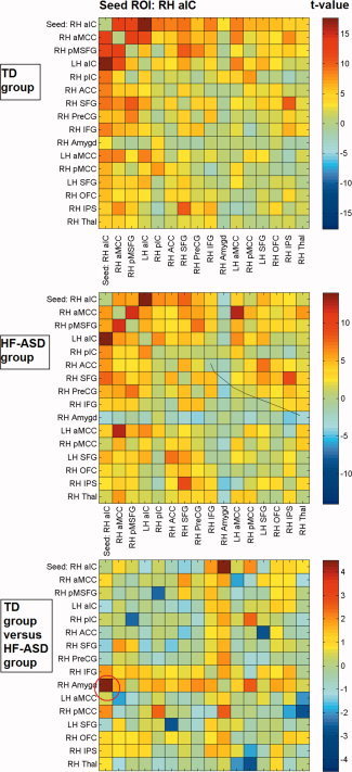
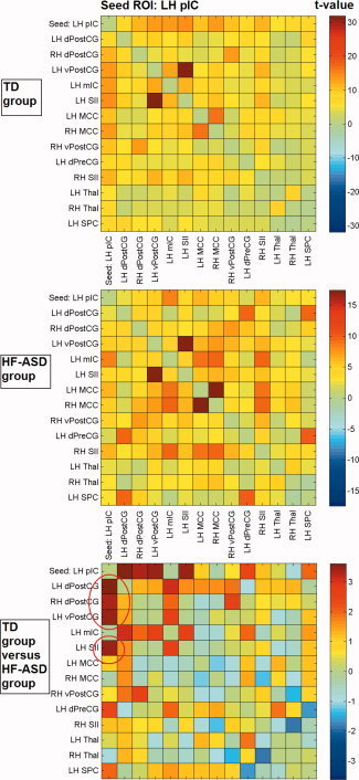
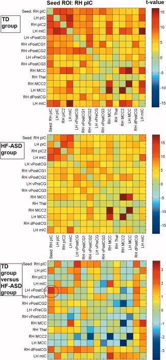
Impaired understanding of others' sensations and emotions as well as abnormal experience of their own emotions and sensations is frequently reported in individuals with Autism Spectrum Disorder (ASD). It is hypothesized that these abnormalities are based on altered connectivity within "shared" neural networks involved in emotional awareness of self and others. The insula is considered a central brain region in a network underlying these functions, being located at the transition of information about bodily arousal and the physiological state of the body to subjective feelings. The present study investigated the intrinsic functional connectivity properties of the insula in 14 high-functioning participants with ASD (HF-ASD) and 15 typically developing (TD) participants in the age range between 12 and 20 years by means of "resting state" or "nontask" functional magnetic resonance imaging. Essentially, a distinction was made between anterior and posterior regions of the insular cortex. The results show a reduced functional connectivity in the HF-ASD group, compared with the TD group, between anterior as well as posterior insula and specific brain regions involved in emotional and sensory processing. It is suggested that functional abnormalities in a network involved in emotional and interoceptive awareness might be at the basis of altered emotional experiences and impaired social abilities in ASD, and that these abnormalities are partly based on the intrinsic functional connectivity properties of such a network.
Copyright © 2010 Wiley-Liss, Inc.
Figures
References
-
- Adolphs R, Tranel D, Damasio H, Damasio A ( 1994): Impaired recognition of emotion in facial expressions following bilateral damage to the human amygdala. Nature 372: 669–672. - PubMed
-
- Anderson AK, Phelps EA ( 2001): Lesions of the human amygdala impair enhanced perception of emotionally salient events. Nature 411: 305–309. - PubMed
-
- Auer DP ( 2008): Spontaneous low‐frequency blood oxygenation level‐dependent fluctuations and functional connectivity analysis of the ‘resting’ brain. Magn Reson Imaging 26: 1055–1064. - PubMed
Publication types
MeSH terms
LinkOut - more resources
Full Text Sources
Research Materials
Miscellaneous
