

Architectural and morphological assessment of rat abdominal wall muscles: comparison for use as a human model
- PMID: 20646108
- PMCID: PMC2919595
- DOI: 10.1111/j.1469-7580.2010.01271.x
Architectural and morphological assessment of rat abdominal wall muscles: comparison for use as a human model
Abstract
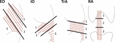
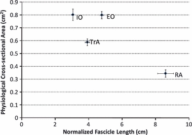
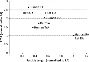
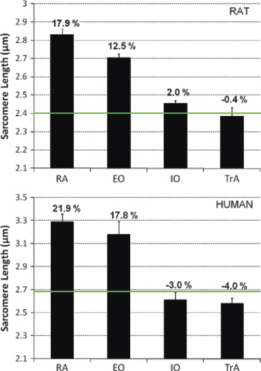
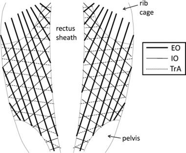
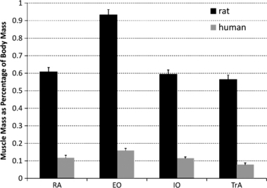
The abdominal wall is a composite of muscles that are important for the mechanical stability of the spine and pelvis. Tremendous clinical attention is given to these muscles, yet little is known about how they function in isolation or how they interact with one another. Given the morphological, vascular, and innervation complexities associated with these muscles and their proximity to the internal organs, an appropriate animal model is important for understanding their physiological and mechanical significance during function. To determine the extent to which the rat abdominal wall resembles that of human, 10 adult male Sprague-Dawley rats were killed and formalin-fixed for architectural and morphological analyses of the four abdominal wall muscles (rectus abdominis, external oblique, internal oblique, and transversus abdominis). Physiological cross-sectional areas and optimal fascicle lengths demonstrated a pattern that was similar to human abdominal wall muscles. In addition, sarcomere lengths measured in the neutral spine posture were similar to human in their relation to optimal sarcomere length. These data indicate that the force-generating and length change capabilities of these muscles, relative to one another, are similar in rat and human. Finally, the fiber lines of action of each abdominal muscle were similar to human over most of the abdominal wall. The main exception was in the lower abdominal region (inferior to the pelvic crest), where the external oblique becomes aponeurotic in human but continues as muscle fibers into its pelvic insertion in the rat. We conclude that, based on the morphology and architecture of the abdominal wall muscles, the adult male Sprague-Dawley rat is a good candidate for a model representation of human, particularly in the middle and upper abdominal wall regions.
Figures
Similar articles
-
Transmission of muscularly generated force and stiffness between layers of the rat abdominal wall.Spine (Phila Pa 1976). 2009 Jan 15;34(2):E70-5. doi: 10.1097/BRS.0b013e31818bd6b1. Spine (Phila Pa 1976). 2009. PMID: 19139656
-
Passive mechanical properties of rat abdominal wall muscles suggest an important role of the extracellular connective tissue matrix.J Orthop Res. 2012 Aug;30(8):1321-6. doi: 10.1002/jor.22068. Epub 2012 Jan 20. J Orthop Res. 2012. PMID: 22267257 Free PMC article.
-
An MRI investigation into the function of the transversus abdominis muscle during "drawing-in" of the abdominal wall.Spine (Phila Pa 1976). 2006 Mar 15;31(6):E175-8. doi: 10.1097/01.brs.0000202740.86338.df. Spine (Phila Pa 1976). 2006. PMID: 16540858
-
Architectural analysis of human abdominal wall muscles: implications for mechanical function.Spine (Phila Pa 1976). 2011 Mar 1;36(5):355-62. doi: 10.1097/BRS.0b013e3181d12ed7. Spine (Phila Pa 1976). 2011. PMID: 21325932 Free PMC article.
-
Mechanically relevant consequences of the composite laminate-like design of the abdominal wall muscles and connective tissues.Med Eng Phys. 2012 May;34(4):521-3. doi: 10.1016/j.medengphy.2011.11.008. Epub 2011 Dec 3. Med Eng Phys. 2012. PMID: 22137674
Cited by
-
Paraspinal muscle pathophysiology associated with low back pain and spine degenerative disorders.JOR Spine. 2021 Sep 15;4(3):e1171. doi: 10.1002/jsp2.1171. eCollection 2021 Sep. JOR Spine. 2021. PMID: 34611593 Free PMC article. Review.
-
A new rat model for orthotopic abdominal wall allotransplantation.Plast Reconstr Surg Glob Open. 2014 May 7;2(4):e136. doi: 10.1097/GOX.0000000000000086. eCollection 2014 Apr. Plast Reconstr Surg Glob Open. 2014. PMID: 25289329 Free PMC article.
-
A Model of Free Tissue Transfer: The Rat Epigastric Free Flap.J Vis Exp. 2017 Jan 15;(119):55281. doi: 10.3791/55281. J Vis Exp. 2017. PMID: 28117814 Free PMC article.
-
A reproducible rat model for predicting incisional hernia recurrence: insights for clinical translation.Sci Rep. 2025 Jul 4;15(1):23861. doi: 10.1038/s41598-025-05557-1. Sci Rep. 2025. PMID: 40615405 Free PMC article.
-
Alterations in the structural characteristics of rectus abdominis muscles caused by diabetes and pregnancy: A comparative study of the rat model and women.PLoS One. 2020 Apr 3;15(4):e0231096. doi: 10.1371/journal.pone.0231096. eCollection 2020. PLoS One. 2020. PMID: 32243473 Free PMC article.
References
-
- Anders C, Wagner H, Puta C, et al. Trunk muscle activation patterns during walking at different speeds. J Electromyogr Kinesiol. 2007;17:245–252. - PubMed
-
- Arjmand N, Shirazi-Adl A, Parnianpour M. Trunk biomechanics during maximum isometric axial torque exertions in upright standing. Clin Biomech. 2008;23:969–978. - PubMed
-
- Arnold JS, Thomas AJ, Kelsen SG. Length-tension relationship of abdominal expiratory muscles: effect of emphysema. J Appl Physiol. 1987;62:739–745. - PubMed
-
- Bodine SC, Roy RR, Meadows DA, et al. Architectural, histochemical, and contractile characteristics of a unique biarticular muscle: the cat semitendinosus. J Neurophysiol. 1982;48:192–201. - PubMed
-
- Brown SHM, McGill SM. Transmission of muscularly generated force and stiffness between layers of the rat abdominal wall. Spine. 2009;34:E70–E75. - PubMed
Publication types
MeSH terms
Grants and funding
LinkOut - more resources
Full Text Sources
