

Epicardium-derived cells (EPDCs) in development, cardiac disease and repair of ischemia
- PMID: 20646126
- PMCID: PMC3822740
- DOI: 10.1111/j.1582-4934.2010.01077.x
Epicardium-derived cells (EPDCs) in development, cardiac disease and repair of ischemia
Abstract
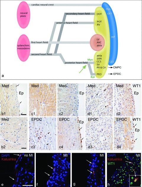
Reactivation of endogenous epicardium after ischemia The proepicardial-derived epicardium covers the myocardium and after a process of epithelial-mesenchymal transition (EMT) forms epicardium-derived cells (EPDCs). These cells migrate into the myocardium and show an essential role in the induction of the ventricular compact myocardium and the differentiation of the Purkinje fibres. EPDCs are furthermore the source of the interstitial fibroblast, the coronary smooth muscle cell and the adventitial fibroblast. The possible differentiation into cardiomyocytes, endothelial cells and the recently described telocyte and other cells in the cardiac stem cell niche needs further investigation. Surgically or genetically disturbed epicardial and EPDC differentiation leads to a spectrum of abnormalities varying from thin undifferentiated myocardium, which can be embryonic lethal, to a diminished coronary vascular bed with even absent main coronary arteries. The embryonic potential of EPDCs has been translated to both structural and functional congenital malformations and adult cardiac disease, like development of Ebstein's malformation, arrhythmia and cardiomyopathies. Furthermore, the use of adult EPDCs as a stem cell source has been explored, showing in an animal model of myocardial ischemia the recapitulation of the embryonic program with improved function, angiogenesis and less adverse remodeling. Combining EPDCs and adult cardiomyocyte progenitor cells synergistically improved these results. The contribution of injected EPDCs was instructive rather than constructive. The finding of reactivation of the endogenous epicardium in ischemia with re-expression of developmental genes and renewed EMT marks the onset of a novel therapeutic focus.
Figures
References
-
- Vrancken Peeters M-PFM, Mentink MMT, Poelmann RE, et al. Cytokeratins as a marker for epicardial formation in the quail embryo. Anat Embryol. 1995;191:503–8. - PubMed
-
- Yang JT, Rayburn H, Hynes RO. Cell adhesion events mediated by alpha 4 integrins are essential in placental and cardiac development. Development. 1995;121:549–60. - PubMed
-
- Mahtab EA, Wijffels MC, Van Den Akker NM, et al. Cardiac malformations and myocardial abnormalities in podoplanin knockout mouse embryos: correlation with abnormal epicardial development. Dev Dyn. 2008;237:847–57. - PubMed
Publication types
MeSH terms
LinkOut - more resources
Full Text Sources
Other Literature Sources
