

Review
doi: 10.1111/j.1365-2826.2010.02021.x.
How it's made: organisational effects of hormones on the developing brain
Affiliations
- PMID: 20646174
- PMCID: PMC3170038
- DOI: 10.1111/j.1365-2826.2010.02021.x
Item in Clipboard
Review
How it's made: organisational effects of hormones on the developing brain
J Neuroendocrinol.
2010 Jul.
Abstract
Gonadal steroid hormones exert permanent organisational effects on the developing brain and thereby direct adult hormonal responsiveness to dictate sex-specific behaviour and physiology. Considerable progress has been made in elucidating the cellular mechanism of action of androgens and oestrogens during the perinatal sensitive period during which organisation occurs. This review highlights the findings obtained with respect to differential cell death and synaptogenesis with an emphasis on region-specific mechanisms that involve diverse signalling molecules including tumour necrosis factor-alpha, BAX, GABA, glutamate, prostaglandin E(2), FAK and paxillin.
Figures
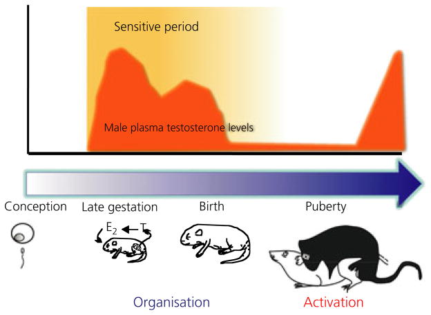
Hormonally-mediated sexual differentiation of the brain. The organisational/activational construct for sexual differentiation is based on the principle of an early sensitive period that is operationally defined as beginning with the onset of gonadal secretion of steroid hormones in the male, and as termination being that point at which the female becomes unresponsive to the impact of exogenously administered steroid hormone. The actions of gonadal steroids during this sensitive period are considered organisational, and generally permanent, and this differentiated neural substrate is then acted upon (activational) by the appropriate gonadal steroids in adulthood to produce stereotypic sexual behaviour or physiology. Different physiological and behavioural endpoints vary in the precise timing of the sensitive period, although most differentiation is complete by the end of the first postnatal week of life. E2, oestradiol.
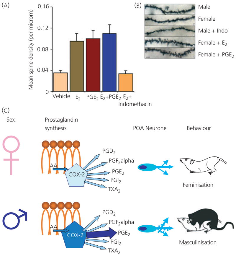
Prostaglandin E2 mediates masculinisation of preoptic area (POA) neurones and adult sexual behaviour of rats. The density of dendritic spines, and the excitatory synapses associated with these structures, is two- to three-fold higher on POA neurones in males than females. Oestradiol permanently organises this sex difference during the perinatal sensitive period. (A) Treatment of females with oestradiol (E2) or prostaglandin E2 (PGE2) significantly increases the density of dendritic spines, to the same level as seen in males. The effects of E2 and PGE2 are not additive, and the effects of E2 are completely blocked by the co-administration of the COX-2 inhibitor, indomethacin (indo). COX-2 is a key enzyme in prostaglandin synthesis (38). (B) Golgi impregnation of POA neurones further reveals the sex difference in dendritic spines and regulation by prostaglandins. These neurones were visualised in 20-day-old animals that had been treated with oestradiol, PGE2 or indomethacin on days 1 and 2 of life. The higher density of spines in males compared to females is evident, as is the induction of spines by E2 or PGE2 treatment of females. Treatment of males with indomethacin permanently down-regulated dendritic spines on POA neurones (16). (C) Schematic representation of the role of prostaglandins in masculinisation. Males have higher levels of the COX-2 enzyme, resulting in five-fold higher levels of the prostaglandin, PGE2, but not other prostanoids. Through a mechanism that at least partly involves the AMPA receptor, but that remains poorly understood, PGE2 permanently increases the density of dendritic spines on POA neurones. The resulting sex difference in neuronal morphology is correlated with (and presumably determines) adult sex differences in reproductive behaviour. However, the changes in brain regions outside the POA contributing to the change in behaviour cannot be ruled out.
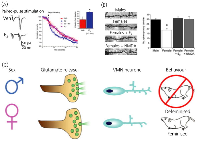
Nongenomic steroid-induced organisation of the hypothalamus. Oestradiol rapidly induces glutamate release from hypothalamic neurones leading to activation of post-synaptic glutamate receptors and induction of dendritic spines. (A) Paired pulse stimulation ratios indicate enhanced release of glutamate on the second stimulation in neurones pretreated with oestradiol. The rate of destaining of FM4-64 visualised terminals confirms this, and the effect of oestradiol is blocked by the highly specific oestrogen receptor antagonist, ICI 182,780. The effect of oestradiol on FM4-64 destaining is apparent within 1 h of treatment and does not require protein synthesis but does require activation of phosphoinositide 3-kinase (PI3) kinase. (B) Golgi impregnated ventromedial nucleus (VMN) neurones reveal a higher number of dendritic spines on the neurones of males or females treated with oestradiol compared to control females (*P < 0.05 compared to all other groups, ANOVA). Treatment with the glutamate agonist, NMDA, mimics the effect of oestradiol, confirming an essential role for glutamate transmission in establishing sexually dimorphic dendritic morphology (25). (C) Schematic representation of the role of glutamate in defeminisation. Enhanced glutamate release in male neurones results in increased dendritic spines, which is correlated with a loss of expression of female sexual behaviour expression in adulthood. Treatment of neonatal females with NMDA successfully defeminises their adult behaviour, further confirming the role of glutamate in the organisational component of sexual differentiation.
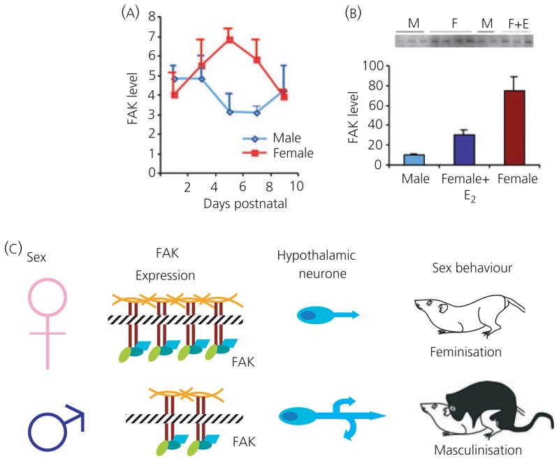
Oestradiol down-regulates focal adhesion kinase (FAK) in the developing male brain. FAK is an important regulator of axonal growth and dendritic branching in the developing brain. Neurones of the developing hypothalamus have more dendritic branches in males than in females, an effect mediated by the higher levels of oestradiol in neonatal male brain, following its aromatisation from testicular androgens. (A) The level of FAK protein gradually increases in the female hypothalamus during the sensitive period of sexual differentiation, peaking on postnatal day 6, at which point it is significantly higher than in the male hypothalamus, where levels remain relatively constant. (B) Treatment of newborn females with oestradiol significantly reduces FAK levels within 24–48 h when assessed by quantitative western blot (28). (C) Schematic representation of FAK, which interacts with the integrins to control cell adhesion, thereby impacting on growth cone movement and branching. Increased FAK levels in females are speculated to decrease branching of hypothalamic neurones. The resulting sex difference in neuronal morphology is considered as a permanently organised change that is then activated by adult circulating gonadal steroids to regulate sexually dimorphic reproductive behaviour.
References
-
- Morris JA, Jordan CL, Breedlove SM. Sexual differentiation of the vertebrate nervous system. Nat Neurosci. 2004;7:1034–1039. - PubMed
-
- Wallen K. Hormonal influences on sexually differentiated behavior in nonhuman primates. Front Neuroendocrinol. 2005;26:7–26. - PubMed
-
- Breedlove SM. Sexual differentiation of the human nervous system. Annu Rev Psychol. 1994;45:389–418. - PubMed
-
- Hines M. Sexual differentiation of human brain and behavior. In: Pfaff D, editor. Hormones, Brain and Behavior. London: Academic Press; 2002. pp. 425–462.
-
- Phoenix CH, Goy RW, Gerall AA, Young WC. Organizing action of prenatally administered testosterone proprionate on the tissues mediating mating behavior in the female guinea pig. Endocrinology. 1959;65:369–382. - PubMed
Publication types
MeSH terms
Substances
Grants and funding
LinkOut - more resources
Full Text Sources
Research Materials
Miscellaneous
