From nose to brain: development of gonadotrophin-releasing hormone-1 neurones
- PMID: 20646175
- PMCID: PMC2919238
- DOI: 10.1111/j.1365-2826.2010.02034.x
From nose to brain: development of gonadotrophin-releasing hormone-1 neurones
Abstract
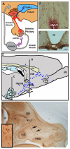
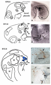
Gonadotrophin-releasing hormone-1 (GnRH-1) is essential for mammalian reproduction, controlling release of gonadotrophins from the anterior pituitary. GnRH-1 neurones migrate from the nasal placode into the forebrain during development. Although first located within the nasal placode, the embryonic origin/lineage of GnRH-1 neurones is still unclear. The migration of GnRH-1 cells is the best characterised example of neurophilic/axophilic migration, with the cells using a subset of olfactory-derived vomeronasal axons as their pathway and numerous molecules to guide their movement into the forebrain. Exciting work in this area is beginning to identify intersecting pathways that orchestrate the movement of these critical neuroendocrine cells into the central nervous system, both spatially and temporally, through a diverse and changing terrain. Once within the forebrain, little is known about how the axons target the median eminence and ultimately secrete GnRH-1 in a pulsatile fashion.
Figures


Similar articles
-
Gonadotropin-releasing hormone neuronal migration.Semin Reprod Med. 2007 Sep;25(5):305-12. doi: 10.1055/s-2007-984736. Semin Reprod Med. 2007. PMID: 17710726 Review.
-
CXCR4/SDF-1 system modulates development of GnRH-1 neurons and the olfactory system.Dev Neurobiol. 2008 Mar;68(4):487-503. doi: 10.1002/dneu.20594. Dev Neurobiol. 2008. PMID: 18188864
-
Dysregulation of Semaphorin7A/β1-integrin signaling leads to defective GnRH-1 cell migration, abnormal gonadal development and altered fertility.Hum Mol Genet. 2011 Dec 15;20(24):4759-74. doi: 10.1093/hmg/ddr403. Epub 2011 Sep 8. Hum Mol Genet. 2011. PMID: 21903667 Free PMC article.
-
Gli3 Regulates Vomeronasal Neurogenesis, Olfactory Ensheathing Cell Formation, and GnRH-1 Neuronal Migration.J Neurosci. 2020 Jan 8;40(2):311-326. doi: 10.1523/JNEUROSCI.1977-19.2019. Epub 2019 Nov 25. J Neurosci. 2020. PMID: 31767679 Free PMC article.
-
Development of gonadotropin-releasing hormone-1 neurons.Front Neuroendocrinol. 2002 Jul;23(3):292-316. doi: 10.1016/s0091-3022(02)00001-8. Front Neuroendocrinol. 2002. PMID: 12127307 Review.
Cited by
-
The Novel Actions of the Metabolite GnRH-(1-5) are Mediated by a G Protein-Coupled Receptor.Front Endocrinol (Lausanne). 2013 Jul 8;4:83. doi: 10.3389/fendo.2013.00083. eCollection 2013. Front Endocrinol (Lausanne). 2013. PMID: 23847594 Free PMC article.
-
Loss-of-function mutations in SOX10 cause Kallmann syndrome with deafness.Am J Hum Genet. 2013 May 2;92(5):707-24. doi: 10.1016/j.ajhg.2013.03.024. Am J Hum Genet. 2013. PMID: 23643381 Free PMC article.
-
Sexually Dimorphic Transcriptomic Changes of Developing Fetal Brain Reveal Signaling Pathways and Marker Genes of Brain Cells in Domestic Pigs.Cells. 2021 Sep 16;10(9):2439. doi: 10.3390/cells10092439. Cells. 2021. PMID: 34572090 Free PMC article.
-
Hypothalamic-pituitary-adrenal and hypothalamic-pituitary-gonadal axes: sex differences in regulation of stress responsivity.Stress. 2017 Sep;20(5):476-494. doi: 10.1080/10253890.2017.1369523. Epub 2017 Aug 31. Stress. 2017. PMID: 28859530 Free PMC article. Review.
-
Illuminating the terminal nerve: Uncovering the link between GnRH-1 neuron and olfactory development.J Comp Neurol. 2024 Mar;532(3):e25599. doi: 10.1002/cne.25599. J Comp Neurol. 2024. PMID: 38488687 Free PMC article.
References
-
- Wray S. Development of gonadotropin-releasing hormone-1 neurons. Frontiers in Neuroendocrinology. 2002;23:292–316. - PubMed
-
- Herbison AE. Physiology of the GnRH neuronal network. In: Neill JD, editor. Knobil and Neill's Physiology of Reproduction. Academic Press; San Diego, CA: 2006. pp. 1415–1482.
-
- Okubo K, Nagahama Y. Structural and functional evolution of gonadotropin-releasing hormone in vertebrates. Acta Physiol (Oxf) 2008;193:3–15. - PubMed
-
- Stewart AJ, Katz AA, Millar RP, Morgan K. Retention and Silencing of PreproGnRH-II and Type II GnRH Receptor Genes in Mammals. Neuroendocrinology. 2009;90:416–432. - PubMed
-
- Palevitch O, Kight K, Abraham E, Wray S, Zohar Y, Gothilf Y. Ontogeny of the GnRH systems in zebrafish brain: in situ hybridization and promoter-reporter expression analyses in intact animals. Cell Tissue Res. 2007;327:313–322. - PubMed
Publication types
MeSH terms
Substances
Grants and funding
LinkOut - more resources
Full Text Sources

