Comparative Study
. 2011 Jan;17(1):77-82.
doi: 10.1111/j.1601-0825.2010.01707.x.

Convenient and reproducible in vivo gene transfer to mouse parotid glands

Affiliations
Comparative Study

Convenient and reproducible in vivo gene transfer to mouse parotid glands

C Zheng et al. Oral Dis. 2011 Jan.

Abstract

Objectives: Published studies of gene transfer to mouse salivary glands have not employed the parotid glands. Parotid glands are the likely target tissue for most clinical applications of salivary gene transfer. The purpose of the present study was to develop a convenient and reproducible method of retroductal gene transfer to mouse parotid glands.

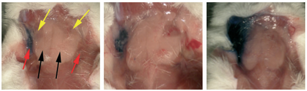
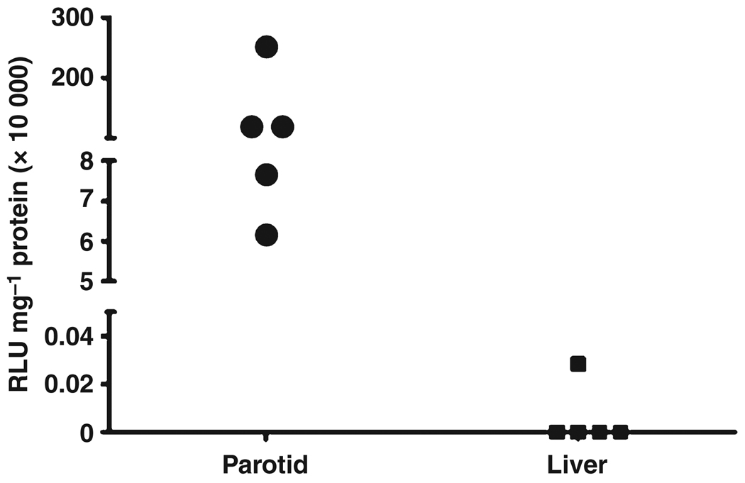
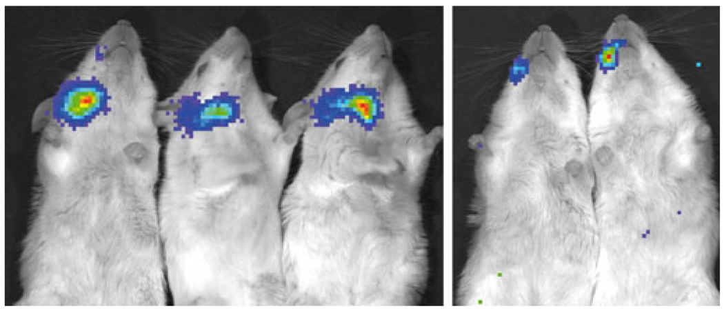
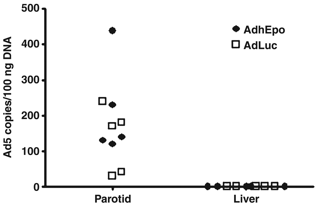
Methods: The volume for vector delivery was assessed by infusion of Toluidine Blue into Stensen's ducts of Balb/c mice after direct intraoral cannulation. Recombinant, serotype 5 adenoviral vectors, encoding either firefly luciferase or human erythropoietin (hEpo), were constructed and then administered to parotid glands (10(7) vector particles/gland). Transgene expression in vivo was measured by enzyme activity (luciferase) or an enzyme-linked immunosorbent assay (hEpo). Vector biodistribution was measured by real-time quantitative (Q) PCR.

Results: The chosen volume for mouse parotid vector delivery was 20μL. Little vector was detected outside of the targeted glands, with both QPCR and luciferase assays. Transgene expression was readily detected in glands (luciferase, hEpo), and serum and saliva (hEpo). Most secreted hEpo was detected in saliva.

Conclusion: These studies show that mouse parotid glands can be conveniently and reproducibly targeted for gene transfer, and should be useful for pre-clinical studies with many murine disease models.


Figures

Figure 1
Figure 1
Determination of fluid volume for use in murine parotid gland vector infusions. The right parotid glands of mice were cannulated and infused with a solution of Toluidine Blue as described in Materials and Methods. The figure shows a mouse infused either with 20- (left), 35- (center) or 50- (right) µl of Toluidine Blue. Red arrows point to parotid glands. Black arrows point to submandibular glands. Yellow arrows point to sublingual glands. No fluid extravasation was seen with the 20-µl volume
Figure 2
Figure 2
Luciferase activity levels achieved following AdLuc delivery to murine parotid glands. Mice were administered AdLuc (107 vector particles) in a single parotid gland. After 48 h mice were sacrificed and parotid and liver tissue was removed. Extracts of tissue were prepared and luciferase activity assayed as described in Materials and Methods. Data shown are individual results from five mice (same animals as shown in Fig. 3)
Figure 3
Figure 3
Visualization of transgene expression in vivo after AdLuc administration to murine parotid glands. AdLuc (107 particles/gland) was delivered to the right parotid glands of five mice and, after 48 h, the tissue distribution of luciferase transgene expression was visualized using the Xenogen IVIS Imaging System. In three mice (left) there were high levels of luciferase activity detected, while in two mice (right) only moderate luciferase activity was detected. No significant luciferase activity can be visualized in any other tissue. See Materials and Methods for additional details
Figure 4
Figure 4
Detection of adenoviral vector in parotid gland and liver tissue of mice following AdLuc or AdhEpo administration. AdLuc or AdhEpo (107 particles/gland) was delivered to single glands (n = 5 mice/vector group), parotid and liver tissue was removed, genomic DNA extracted and QPCR assays performed as described in Materials and Methods. Data from individual mice are shown in a scatter plot (open squares, AdLuc; closed diamonds, AdhEpo)
Figure 5
Figure 5
Morphological evaluation of murine parotid glands following Ad5 vector administration. Parotid glands from control and treated mice were fixed in 10% formalin and then paraffin-embedded. Ten µm sections were stained with either H&E (a, b) or immunostained with a primary antibody directed to hEpo (c, d), see Materials and Methods for additional details. (a, c) Sections from a control parotid gland. (b, d) sections from a parotid gland transduced with AdhEpo

Similar articles

Cited by

References

    1. Adesanya MR, Redman RS, Baum BJ, O’Connell BC. Immediate inflammatory responses to adenovirus-mediated gene transfer in rat salivary glands. Hum Gene Ther. 1996;7:1085–1093. - PubMed
    1. Amalfitano A, McVie-Wylie AJ, Hu H, et al. Systemic correction of the muscle disorder glycogen storage disease type II after hepatic targeting of a modified adenovirus vector encoding human acid-α-glucosidase. Proc Natl Acad Sci USA. 1999;96:8861–8866. - PMC - PubMed
    1. Barka T, van der Noen H. Retrovirus-mediated gene transfer into salivary glands in vivo. Hum Gene Ther. 1996;7:613–618. - PubMed
    1. Baum BJ, Wellner RB, Zheng C. Gene transfer to salivary glands. Int Rev Cytol. 2002;213:93–146. - PubMed
    1. Baum BJ, Zheng C, Alevizos I, et al. Development of a gene transfer-based treatment for radiation-induced salivary hypofunction. Oral Oncol. 2010;46:4–8. - PMC - PubMed

Publication types

MeSH terms