Review
. 2011 Apr 13;51(7):613-32.
doi: 10.1016/j.visres.2010.07.010. Epub 2010 Jul 18.

Development of the retina and optic pathway

Affiliations
Review

Development of the retina and optic pathway

Benjamin E Reese. Vision Res. .

Abstract

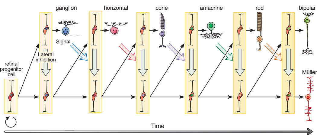
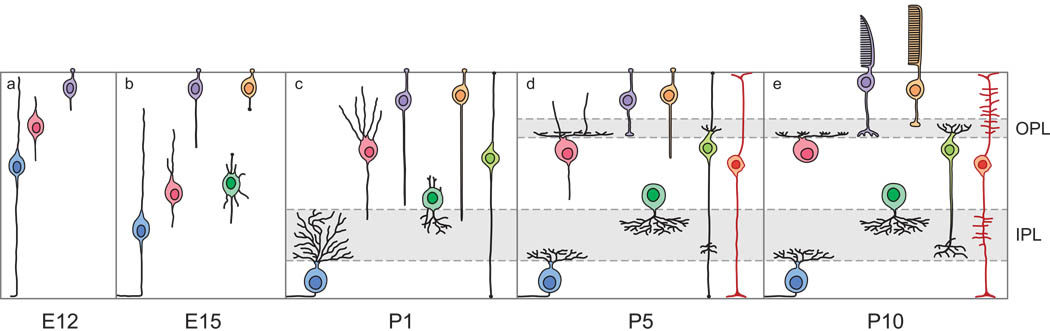
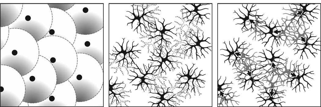
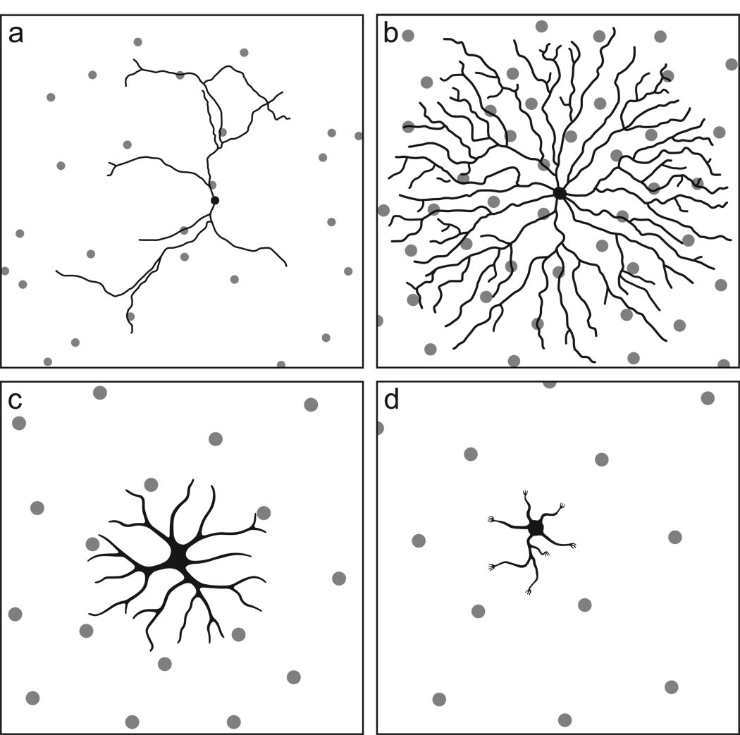
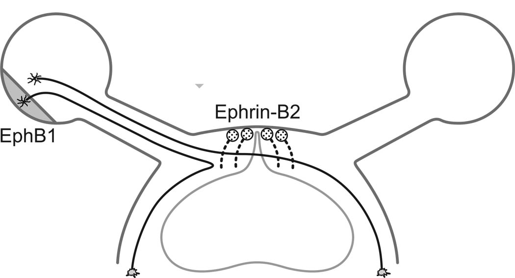
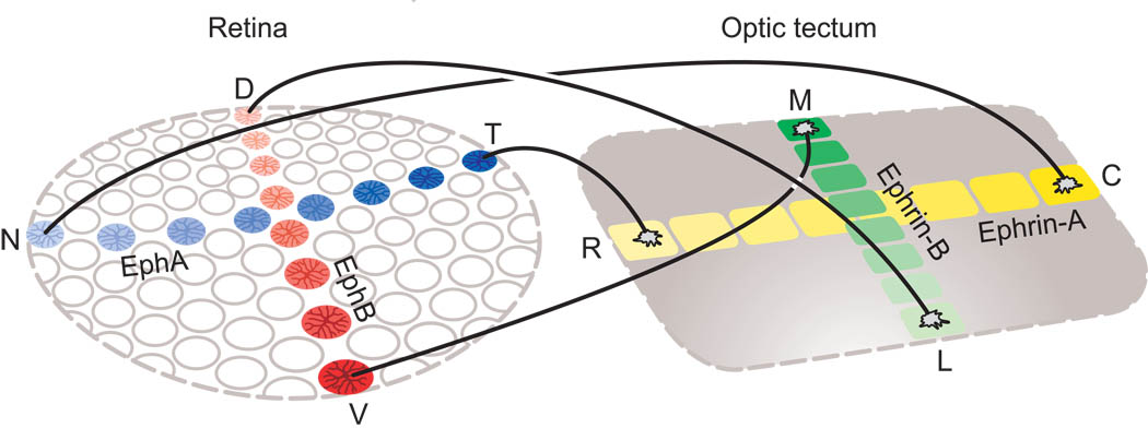
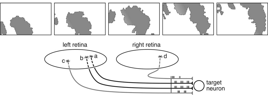
Our understanding of the development of the retina and visual pathways has seen enormous advances during the past 25years. New imaging technologies, coupled with advances in molecular biology, have permitted a fuller appreciation of the histotypical events associated with proliferation, fate determination, migration, differentiation, pathway navigation, target innervation, synaptogenesis and cell death, and in many instances, in understanding the genetic, molecular, cellular and activity-dependent mechanisms underlying those developmental changes. The present review considers those advances associated with the lineal relationships between retinal nerve cells, the production of retinal nerve cell diversity, the migration, patterning and differentiation of different types of retinal nerve cells, the determinants of the decussation pattern at the optic chiasm, the formation of the retinotopic map, and the establishment of ocular domains within the thalamus.


Figures

Figure 1
Figure 1
Retinal progenitor cells are multipotent, competent to produce all types of retinal cell. This competence is progressively restricted, ensuring a unidirectional sequence of cell production, but not all progenitors at the outset of neurogenesis will produce each type of cell. At the left, early symmetrical divisions expand the population of progenitors. As their numbers increase, so do associated signals derived from them, eventually allowing proneural gene expression and Notch-Delta mediated lateral inhibition (vertical grey arrows) to permit only a subset of progenitors to leave the cell cycle (defining the onset of neurogenesis) and to differentiate. Newborn neurons provide signals (diagonal colored arrows) that restrict production of the same cell types, directing later-generated cells to subsequent fates. With the acquisition of additional cell types, so the micro-environment changes progressively (saturating vertical rectangles), and these extrinsic signals, coupled with intrinsic changes, alter the transcription factors expressed by progenitors as a function of time (colored nuclei). Thus changing cellular competence, intercellular signaling between progenitors, and secreted signaling by differentiating cells interact to yield variability in clonal constituency. The schematic implies a stem-like lineage tree, but by the insertion of additional divisions at various locations along this time-line, progenitors within this lineage tree may yield terminal divisions producing two postmitotic daughters (e.g. a ganglion cell and a cone photoreceptor, or a pair of horizontal cells, or a later-generated neuron and Muller glial cell, as shown at the far right), or daughters that both divide again, thereby amplifying the population of progenitors that produce later-generated cell types (e.g. rod photoreceptors and bipolar cells). (Modified from Agathocleous & Harris,2009).
Figure 2
Figure 2
Retinal precursors initiate a cell-type specific differentiation program, controlling their migratory behavior and their morphogenesis. Shown are five different developmental time-points, conveying these migratory and morphological distinctions as the different cell-types achieve their laminar positioning and adult morphologies. Cell type conventions as in figure 1. (Modified from Mumm & Lohmann, 2006).
Figure 3
Figure 3
The regularity of a retinal mosaic may be the product of distinct developmental events acting at different stages, dependent upon cell type. Periodic fate determining events may establish a regular pattern amongst a population of retinal precursors forming a retinal mosaic (a). At later stages during differentiation, naturally occurring cell death may eliminate closely positioned neighbors (dotted profiles in b), because of limited trophic support from afferents or targets. Mutual repulsion may also drive homotypic cells apart (arrows in c). (Modified from Reese, 2008b).
Figure 4
Figure 4
The morphology of four different retinal cell types in relation to their retinal mosaics. a. Dopaminergic amacrine cells differentiate a dendritic arbor that is indifferent to the presence of other dendrites arising from the same cell as well as from those of homotypic neighbors. b. Cholinergic amacrine cells differentiate a dendritic arbor in which dendrites avoid one another, yet overlaps those of numerous homotypic neighbors. c. Horizontal cell somata constrain further dendritic outgrowth from their homotypic neighbors. d. Bipolar cell dendritic arbors achieve a tiling of the retinal surface, colonizing pedicles within their dendritic fields while also sharing those at the dendritic boundary with adjacent cells. The panels are not drawn to the same scale, intending only to portray the relationship between morphology and homotypic density. (Modified from Reese, 2008b).
Figure 5
Figure 5
The transcription factor Islet-2 represses Zic2 expression, which in turn regulates EphB1 receptor expression in the ventro-temporal retina. Ganglion cells in this region of the retina are therefore equipped to detect and respond to the presence of Ephrin-B2 on the surface of a select group of radial glia at the midline, deflecting these optic axons ipsilaterally. (Modified from Petros et al., 2009).
Figure 6
Figure 6
Orthogonal density gradients of EphA3 and EphB2/EphB3 receptors upon the population of retinal ganglion cells enable their axonal growth cones to identify corresponding locations within the optic tectum by virtue of complementary Ephrin-A2/A5 and Ephrin-B1 gradients, mediating the establishment of the retinotopic map.
Figure 7
Figure 7
Spontaneous (pre-visual) waves of activity spread across the surface of the developing retina (top), affecting each retina independently. The top five panels show the active regions (shaded) at two-second intervals from left to right. As a consequence, the bursts of action potentials by retinal ganglion cells will be more highly correlated between those axon terminals arising from adjacent locations on the retinal surface (a and b), ensuring a stabilization of their nascent synaptic contacts with target neurons, refining the precision of the retinotopic map (bottom). Much as more distant locations on the retina will have non-synchronous spontaneous activity with these two cells (c), so locations on the opposite retina should generate asynchronous discharges (d) relative to these cells, leading to the loss of binocular innervation upon single cells in the developing lateral geniculate nucleus. (Top modified from Stafford, Sher, Litke, & Feldheim, 2009).

References

    1. Adler R. A model of retinal cell differentiation in the chick embryo. Prog. Ret. Eye Res. 2000;19:529–557. - PubMed
    1. Agathocleous M, Harris WA. Cell determination. In: Sernagor E, Eglen S, Harris B, Wong R, editors. Retinal Development. Cambridge: Cambridge University Press; 2006. pp. 75–98.
    1. Agathocleous M, Harris WA. From progenitors to differentiated cells in the vertebrate retina. Ann. Rev. Cell Develop. Biol. 2009;25:45–69. - PubMed
    1. Agathocleous M, Iordanova I, Willardsen MI, Xue XY, Vetter ML, Harris WA, Moore KB. A directional Wnt/beta-catenin-Sox2-proneural pathway regulates the transition from proliferation to differentiation in the Xenopus retina. Development. 2009;136:3289–3299. - PMC - PubMed
    1. Ajioka I, Martins RAP, Bayazitov IT, Donovan S, Johnson DA, Frase S, Cicero SA, Boyd K, Zakharenko SS, Dyer MA. Differentiated horizontal interneurons clonally expand to form metastatic retinoblastoma in mice. Cell. 2007;131:378–390. - PMC - PubMed

Publication types