. 2010 Oct 1;34(7):1285-93.
doi: 10.1016/j.pnpbp.2010.07.012. Epub 2010 Jul 18.

Multiple anxiogenic drugs recruit a parvalbumin-containing subpopulation of GABAergic interneurons in the basolateral amygdala

Affiliations

Multiple anxiogenic drugs recruit a parvalbumin-containing subpopulation of GABAergic interneurons in the basolateral amygdala

Matthew W Hale et al. Prog Neuropsychopharmacol Biol Psychiatry. .

Abstract

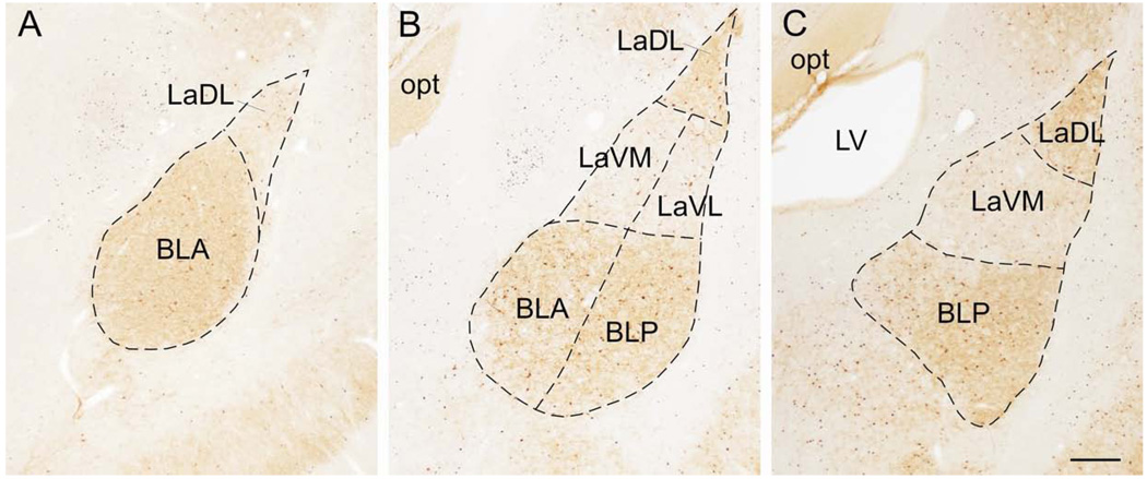
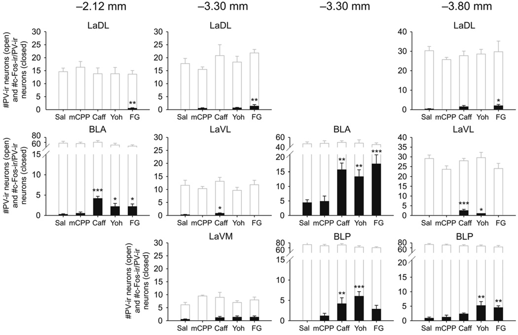
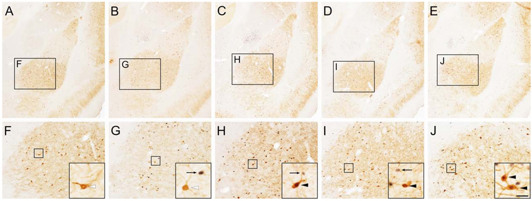
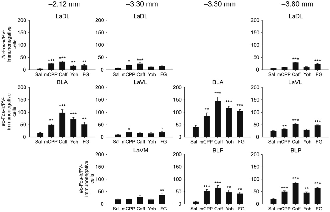
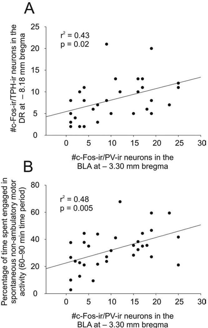
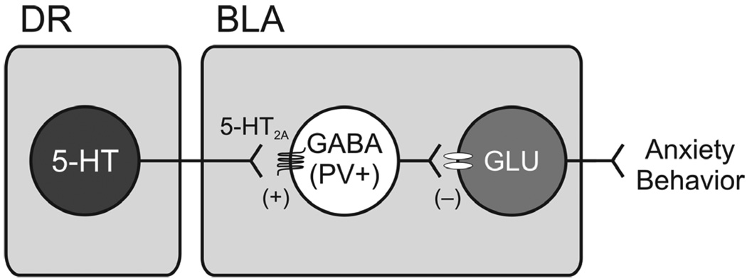
The basolateral amygdala is a nodal structure within a distributed and interconnected network that regulates anxiety states and anxiety-related behavior. Administration of multiple anxiogenic drugs increases cellular responses (i.e., increases c-Fos expression) in a subregion of the basolateral amygdala, but the neurochemical phenotypes of these cells are not known. The basolateral amygdala contains glutamatergic projection neurons and several populations of γ-aminobutyric acid-synthesizing (GABAergic) interneurons, including a population of parvalbumin (PV)-expressing GABAergic interneurons that co-express the excitatory 5-HT(2A) receptor. The role for these PV-expressing GABAergic interneurons in anxiety-states is unclear. In this experiment we examined the effects of multiple anxiogenic drugs including the 5-HT(2C/2A) receptor agonist m-chlorophenyl piperazine (mCPP), the adenosine receptor antagonist caffeine, the α(2)-adrenoreceptor antagonist yohimbine and the partial inverse agonist at the benzodiazepine allosteric site on the GABA(A) receptor, N-methyl-beta-carboline-3-carboxamide (FG-7142), on c-Fos expression in PV-immunoreactive (PV-ir) interneurons in subdivisions of the basolateral amygdala. All drugs with the exception of mCPP increased c-Fos expression in PV-ir neurons in the basolateral amygdaloid nucleus, anterior part (BLA). The numbers of c-Fos-immunoreactive (c-Fos-ir)/PV-ir GABAergic interneurons in the BLA were positively correlated with the numbers of c-Fos-ir serotonergic neurons in the mid-rostrocaudal dorsal raphe nucleus (DR) and with a measure of anxiety-related behavior. All four drugs increased c-Fos expression in non-PV-ir cells in most of the subdivisions of the basolateral amygdala that were sampled, compared with vehicle-injected controls. Together, these data suggest that the PV/5-HT(2A) receptor expressing GABAergic interneurons in the basolateral amygdala are part of a DR-basolateral amygdala neuronal circuit modulating anxiety-states and anxiety-related behavior.


Figures

Figure 1
Figure 1
Photomicrographs showing parvalbumin (PV) and c-Fos immunostaining in the subregions of the basolateral amygdaloid complex at −2.12 mm, −3.30 mm and −3.80 mm from bregma. A) Low power photomicrograph showing the basolateral amygdaloid complex at approximately −2.12 mm bregma. Dashed lines show the borders of the subregions including the lateral amygdaloid nucleus, dorsolateral part (LaDL) and basolateral amygdaloid nucleus, anterior part (BLA). B) Low power photomicrograph showing the basolateral amygdaloid complex at approximately −3.30 mm bregma. Dashed lines show the borders of the subregions including the LaDL, lateral amygdaloid nucleus, ventrolateral part (LaVL), lateral amygdaloid nucleus, ventromedial part, BLA, and basolateral amygdaloid nucleus, posterior part (BLP). C) Low power photomicrograph showing the basolateral amygdaloid complex at approximately −3.80 mm bregma. Dashed lines show the borders of the subregions including the LaDL, LaVM and BLP. Abbreviations: LV, lateral ventricle; opt, optic tract. Scale bar, 250 µm.
Figure 2
Figure 2
Anxiogenic drugs increased c-Fos expression in parvalbumin (PV)-immunoreactive (ir) neurons in the basolateral amygdaloid nucleus complex. Graphs show the numbers of c-Fos-ir/PV-ir neurons and total numbers of PV-ir neurons in subdivisions of the basolateral amygdaloid complex at −2.12 mm bregma (left column), −3.30 mm bregma (middle columns) and −3.80 mm bregma (right column). Closed bars represent the numbers of c-Fos-immunoreactive (c-Fos-ir)/PV-ir neurons. Open bars represent the total numbers of PV-ir neurons within each subdivision. *p < 0.05, **p < 0.01, ***p < 0.001 versus saline-treated controls, post hoc Dunnett’s tests, (saline, n = 8; mCPP, n = 6; caffeine, n = 6; yohimbine, n = 6; FG-7142, n = 6). Abbreviations: BLA, basolateral amygdaloid nucleus, anterior part; BLP, basolateral amygdaloid nucleus, posterior part; Caff, caffeine; FG, N-methyl-beta-carboline-3-carboxamide (FG-7142); LaDL, lateral amygdaloid nucleus, dorsolateral part; LaVL, lateral amygdaloid nucleus, ventrolateral part; LaVM, lateral amygdaloid nucleus, ventromedial part; mCPP, m-chlorophenyl piperazine; Sal, saline; Yoh, yohimbine.
Figure 3
Figure 3
Photomicrographs illustrating c-Fos expression in parvalbumin (PV)-immunoreactive (ir) neurons in the basolateral amygdaloid nucleus, anterior part (BLA) at approximately −3.30 mm bregma in rats injected with A) saline; B) m-chlorophenyl piperazine (mCPP); C) caffeine; D) yohimbine; and E) N-methyl-beta-carboline-3-carboxamide (FG-7142). A–E) Low power photomicrographs showing the basolateral amygdaloid complex. Black boxes in A–E are shown at higher magnification in F–J. F–J) Higher power photomicrographs showing the basolateral amygdaloid nucleus. Black boxes in F–J are shown at higher magnification in insets in the lower right corner of each panel. Black arrows indicate c-Fos-ir/PV-immunonegative cells (blue/black nuclear staining), white arrowheads indicate c-Fos-immunonegative/PV-ir neurons (brown/orange cytoplasmic staining) and black arrowheads indicate c-Fos-ir/PV-ir (double-immunostained) neurons. Scale bar, 250 µm (A–E), 100 µm (F–J), 25 µm (insets).
Figure 4
Figure 4
Anxiogenic drugs increased c-Fos expression in non-parvalbumin (PV)-immunoreactive (ir) cells in the basolateral amygdaloid complex. Graphs show the numbers of c-Fos-ir/PV-immunonegative cells in subdivisions of the basolateral amygdaloid complex at −2.12 mm bregma (left column), −3.30 mm bregma (middle columns) and −3.80 mm bregma (right column). *p < 0.05, **p < 0.01, ***p < 0.001 versus saline-treated controls, post hoc Dunnett’s tests, (saline, n = 8; mCPP, n = 6; caffeine, n = 6; yohimbine, n = 6; FG-7142, n = 6). For abbreviations see Figure 2 legend.
Figure 5
Figure 5
Activation of parvalbumin (PV)-expressing γ-aminobutyric acid (GABA)ergic interneurons in the basolateral amygdaloid nucleus, anterior part (BLA) was associated with activation of serotonergic neurons in the dorsal raphe nucleus, dorsal part (DR) and anxiety-related behavior. A) Scatter and line plot showing a positive correlation between the numbers of c-Fos-immunoreactive (ir)/PV-ir neurons in the BLA and the numbers of c-Fos-ir/tryptophan hydroxylase-ir neurons in the dorsal raphe nucleus (DR) at −8.18 mm bregma. B) Scatter and line plot showing a positive correlation between the numbers of c-Fos-ir/PV-ir neurons in the BLA and the percentage of time spent engaged in spontaneous non-ambulatory motor activity (SNAMA) in the 60–90 min time period post drug injection.
Figure 6
Figure 6
Hypothetical model in which activation of dorsal raphe nucleus (DR) serotonergic neurons results in inhibition of projection neurons within the basolateral amygdala, anterior part (BLA) via activation of the 5-HT2A receptor-expressing subpopulation of local parvalbumin (PV)-expressing γ-aminobutyric acid (GABA)ergic interneurons, resulting in a reduction of excitatory output to circuits mediating anxiety-related behavior.

Publication types

MeSH terms