Review
. 2011 Mar;1810(3):339-49.
doi: 10.1016/j.bbagen.2010.07.005. Epub 2010 Jul 18.

Protein-engineered biomaterials: nanoscale mimics of the extracellular matrix

Affiliations
Review

Protein-engineered biomaterials: nanoscale mimics of the extracellular matrix

Nicole H Romano et al. Biochim Biophys Acta. 2011 Mar.

Abstract

Background: Traditional materials used as in vitro cell culture substrates are rigid and flat surfaces that lack the exquisite nano- and micro-scale features of the in vivo extracellular environment. While these surfaces can be coated with harvested extracellular matrix (ECM) proteins to partially recapitulate the bio-instructive nature of the ECM, these harvested proteins often exhibit large batch-to-batch variability and can be difficult to customize for specific biological studies. In contrast, recombinant protein technology can be utilized to synthesize families of 3 dimensional protein-engineered biomaterials that are cyto-compatible, reproducible, and fully customizable.

Scope of review: Here we describe a modular design strategy to synthesize protein-engineered biomaterials that fuse together multiple repeats of nanoscale peptide design motifs into full-length engineered ECM mimics.

Major conclusions: Due to the molecular-level precision of recombinant protein synthesis, these biomaterials can be tailored to include a variety of bio-instructional ligands at specified densities, to exhibit mechanical properties that match those of native tissue, and to include proteolytic target sites that enable cell-triggered scaffold remodeling. Furthermore, these biomaterials can be processed into forms that are injectable for minimally-invasive delivery or spatially patterned to enable the release of multiple drugs with distinct release kinetics.

General significance: Given the reproducibility and flexibility of these protein-engineered biomaterials, they are ideal substrates for reductionist biological studies of cell-matrix interactions, for in vitro models of physiological processes, and for bio-instructive scaffolds in regenerative medicine therapies. This article is part of a Special Issue entitled Nanotechnologies - Emerging Applications in Biomedicine.


Figures

Figure 1
Figure 1
Schematic of the modular design strategy used to create two families of protein-engineered biomaterials. Top: A chemically-crosslinked hydrogel fabricated from multiple repeats of elastin-like modules, cell adhesion modules, and protease degradation modules [6, 37]. The engineered proteins form a chemical hydrogel network through covalent bonding between a crosslinker and multiple lysine amino acid residues on neighboring protein chains. Bottom: A mixing-induced two-component hydrogel fabricated from two repeating peptide sequences that hetero-assemble [14]. The engineered proteins form a physical hydrogel network through transient hydrogen bonding between WW domains and PPxY domains on neighboring protein chains.
Figure 2
Figure 2
Flow chart showing the sequence of experimental steps used to fabricate a protein-engineered biomaterial. Once a sequence of repetitive peptide modules is designed, the primary amino acid sequence is encoded in a recombinant gene. Solid-state oligonucleotide synthesis and molecular biology cloning are used to create a plasmid harboring the recombinant gene. The plasmid is transfected into the host of choice, often Escherichia coli. The biosynthetic machinery of the host translates the genetic message into an expressed engineered protein. The target protein is purified away from the host contaminants; for example, differential solubility induced by temperature cycling is often used to purify elastin-like proteins [6]. The proteins are processed into a suitable scaffold through chemical or physical crosslinking. The scaffolds can be used for both 2D and 3D cell culture techniques.
Figure 3
Figure 3
Images of an elastin-like, protein-engineered biomaterial. A. Photograph of a chemically crosslinked scaffold, 5 mm in diameter, 2 mm in height. B. Phase contrast micrograph of human umbilical vein endothelial cells (HUVEC) growing within a 3D environment inside a chemically crosslinked scaffold.
Figure 4
Figure 4
Customizing the identity and density of bio-instructional ligands. Top: Encoding various nanoscale ligand modules into the primary amino acid sequence will yield protein-engineered biomaterials that elicit specific functionalities. Bottom: The density of the ligand present in the scaffold can be tailored without altering the overall protein density; therefore, these are ideal scaffolds for reductionist single-variable studies.
Figure 5
Figure 5
Independent customization of ligand density and mechanical properties. Increasing the bio-instructional ligand density (vertical axis) without altering the overall protein density will not affect the mechanical properties of the scaffold. Increasing the crosslinking density (horizontal axis) without altering the overall protein density will increase the scaffold rigidity without affecting the ligand density. Therefore, this strategy is used to create a family of related ECM-mimetic biomaterials with customized properties.
Figure 6
Figure 6
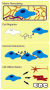
Schematic of physiological processes that can be modeled in vitro using protein-engineered biomaterials. Many physiological processes occur in 3D micro-environments that are difficult to access in vivo and are difficult to accurately recreate in vitro. Protein-engineered biomaterials are suitable scaffolds to be customized for ECM-mimetic in vitro models of cell-matrix remodeling, cell migration, collective cell-cell interactions, and stem/progenitor cell differentiation.
Figure 7
Figure 7
Schematic of drug delivery strategies for protein-engineered biomaterials. The release rate of an encapsulated drug can be customized through selection of an appropriate delivery strategy. The simplest strategy is to physically entrap the drug within the pores of the network and to allow delivery to occur through diffusion. Drug release can be retarded by designing interactions between the drug and network, such as affinity binding, which decrease the effective diffusion rate. By covalently grafting the drug to the protein chain, the drug release can be targeted to occur in response to network degradation, such as protease-induced chain cleavage. Finally, if the drug is a peptide pharmaceutical, the drug can be designed directly into the primary amino acid sequence of the scaffold and released upon network degradation.
Figure 8
Figure 8
Customization of drug release profiles from protein-engineered biomaterials. A. Phase contrast micrograph of a 3D patterned elastin-like biomaterial designed to release two model drugs with two distinct spatial and temporal delivery profiles [57]. Scale bar = 1 mm. Two fluorescently-labeled model drugs were covalently grafted to two different engineered proteins, patterned into two disc-shaped drug depots, and encapsulated within a third engineered protein by chemical crosslinking. The upper drug depot, which is fabricated from a protein designed to slowly degrade in response to the protease uPA, provides a sustained release of the model drug. The lower drug depot, which is fabricated from a protein designed to quickly degrade in response to uPA, provides a burst-like triggered release of the model drug. After several days, the upper drug depot is still present while the lower drug depot has completely disappeared. B. Schematic of three potential drug release profiles that can be designed into protein-engineered biomaterials: burst release, timed burst release (which can be designed to occur in response to a specific biochemical trigger like the protease uPA from the example in panel A), and sustained release.

References

    1. Park H, Cannizzaro C, Vunjak-Novakovic G, Langer R, Vacanti CA, Farokhzad OC. Nanofabrication and microfabrication of functional materials for tissue engineering. Tissue Eng. 2007;13:1867–1877. - PubMed
    1. Szu-Yuan C, Chao-Min C, LeDuc PR. Composite polymer systems with control of local substrate elasticity and their effect on cytoskeletal and morphological characteristics of adherent cells. Biomaterials. 2009;30:3136–3142. - PubMed
    1. Discher DE, Janmey P, Wang YL. Tissue cells feel and respond to the stiffness of their substrate. Science. 2005;310:1139–1143. - PubMed
    1. Chen CS. Mechanotransduction - a field pulling together? J Cell Sci. 2008;121:3285–3292. - PubMed
    1. Wong Po Foo C, Heilshorn SC. Protein-engineered biomaterials. In: Park SJ, Cochran JR, editors. Protein Engineering and Design. CRC Press; Boca Raton: 2010.

Publication types