

The expression patterns of minor fibrillar collagens during development in zebrafish
- PMID: 20647059
- PMCID: PMC2956583
- DOI: 10.1016/j.gep.2010.07.002
The expression patterns of minor fibrillar collagens during development in zebrafish
Abstract
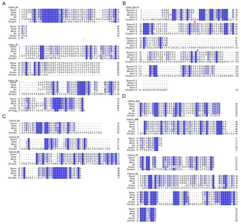
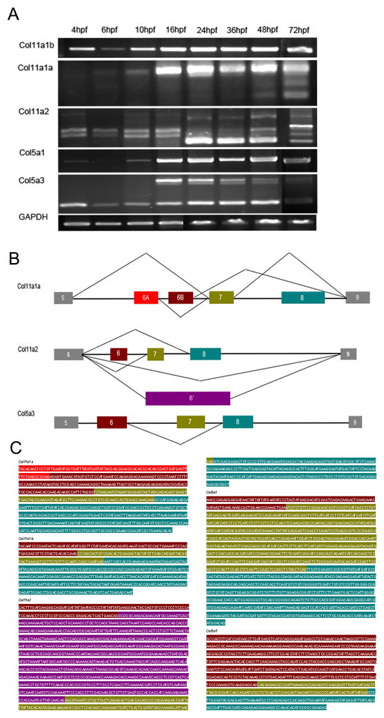
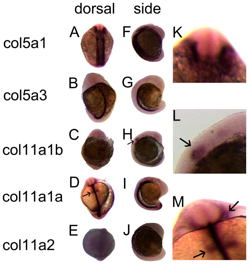
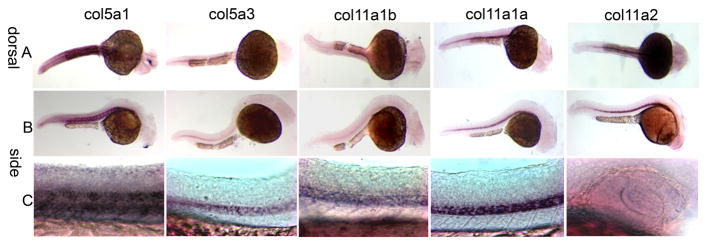
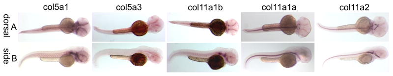
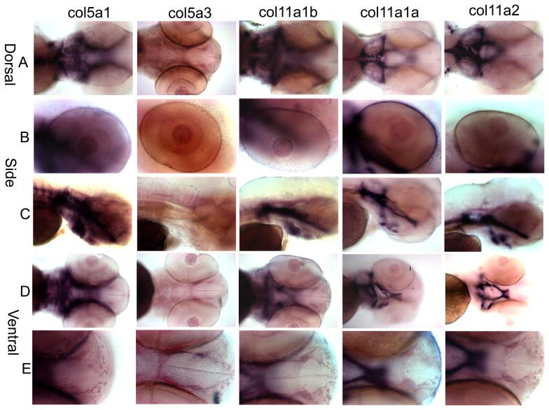
Minor fibrillar collagens are recognized as the organizers and nucleators during collagen fibrillogenesis but likely serve additional functions. The minor fibrillar collagens include collagens type V and XI. Mutations of collagens type V and XI can cause Ehlers-Danlos, Stickler's, and Marshall's syndromes in human. We have characterized the spatiotemporal expression patterns of Col11a1, Col11a2, Col5a1 as well as Col5a3 in zebrafish embryos by in situ hybridization. Col5a1 is expressed in developing somites, neural crest, the head mesenchyme, developing cranial cartilage, pharyngeal arches and vertebrae. Col5a3 is detected in the notochord, mesenchyme cells in the eyes and lens. Both Col11a1 and Col11a2 have similar expression patterns, including notochord, otic vesicle, and developing cranial cartilages. Zebrafish may therefore serve as a valuable vertebrate model system for the study of diseases associated with collagens type V and XI mutations.
Copyright © 2010 Elsevier B.V. All rights reserved.
Figures
Similar articles
-
Craniofacial cartilage morphogenesis requires zebrafish col11a1 activity.Matrix Biol. 2009 Oct;28(8):490-502. doi: 10.1016/j.matbio.2009.07.004. Epub 2009 Jul 26. Matrix Biol. 2009. PMID: 19638309
-
Critical early roles for col27a1a and col27a1b in zebrafish notochord morphogenesis, vertebral mineralization and post-embryonic axial growth.PLoS One. 2009 Dec 29;4(12):e8481. doi: 10.1371/journal.pone.0008481. PLoS One. 2009. PMID: 20041163 Free PMC article.
-
Tgfbeta3 regulation of chondrogenesis and osteogenesis in zebrafish is mediated through formation and survival of a subpopulation of the cranial neural crest.Mech Dev. 2010 Jul-Aug;127(7-8):329-44. doi: 10.1016/j.mod.2010.04.003. Epub 2010 Apr 18. Mech Dev. 2010. PMID: 20406684
-
Molecular Basis of Pathogenic Variants in the Fibrillar Collagens.Genes (Basel). 2022 Jul 4;13(7):1199. doi: 10.3390/genes13071199. Genes (Basel). 2022. PMID: 35885981 Free PMC article. Review.
-
Minor fibrillar collagens, variable regions alternative splicing, intrinsic disorder, and tyrosine sulfation.Protein Cell. 2012 Jun;3(6):419-33. doi: 10.1007/s13238-012-2917-5. Epub 2012 Jul 1. Protein Cell. 2012. PMID: 22752873 Free PMC article. Review.
Cited by
-
The Shape of the Jaw-Zebrafish Col11a1a Regulates Meckel's Cartilage Morphogenesis and Mineralization.J Dev Biol. 2022 Sep 22;10(4):40. doi: 10.3390/jdb10040040. J Dev Biol. 2022. PMID: 36278545 Free PMC article.
-
Col5a3 Likely Promotes Adipogenesis of 3T3-L1 Through Oxidative Phosphorylation.Genes (Basel). 2025 Jan 27;16(2):165. doi: 10.3390/genes16020165. Genes (Basel). 2025. PMID: 40004494 Free PMC article.
-
Authentication of a novel antibody to zebrafish collagen type XI alpha 1 chain (Col11a1a).BMC Res Notes. 2021 Sep 15;14(1):359. doi: 10.1186/s13104-021-05770-x. BMC Res Notes. 2021. PMID: 34526111 Free PMC article.
-
The development of the myotendinous junction. A review.Muscles Ligaments Tendons J. 2012 Sep 10;2(2):53-63. Print 2012 Apr. Muscles Ligaments Tendons J. 2012. PMID: 23738275 Free PMC article.
-
Transcriptome analysis of adipose tissue from pigs divergent in feed efficiency reveals alteration in gene networks related to adipose growth, lipid metabolism, extracellular matrix, and immune response.Mol Genet Genomics. 2019 Apr;294(2):395-408. doi: 10.1007/s00438-018-1515-5. Epub 2018 Nov 27. Mol Genet Genomics. 2019. PMID: 30483895
References
-
- Annunen S, Körkkö J, Czarny M, Warman ML, Brunner HG, Kääriäinen H, Mulliken JB, Tranebjæg L, Brooks DG, Cox GF, Cruysberg JR, Curtis MA, Davenport SLH, Friedrich CA, Kaitila I, Krawczynski MR, Latos-Bielenska A, Mukai S, Olsen BR, Shinno N, Somer M, Vikkula M, Zlotogora J, Prockop DJ, Ala-Kokko L. Splicing mutations of 54-bp exons in the COL11A1 gene cause Marshall syndrome, but other mutations cause overlapping Marshall/Stickler phenotypes. Am J Hum Genet. 1999;65:974–983. - PMC - PubMed
-
- Baas D, Malbouyres M, Haftek-Terreau Z, Le Guellec D, Ruggiero F. Craniofacial cartilage morphogenesis requires zebrafish col11a1 activity. Matrix Biol. 2009;28:490–502. - PubMed
-
- Bisgrove BW, Essner JJ, Yost HJ. Regulation of midline development by antagonism of lefty and nodal signaling. Development. 1999;126:3253–3262. - PubMed
-
- Chernousov MA, Rothblum K, Tyler WA, Stahl RC, Carey DJ. Schwann Cells Synthesize Type V Collagen That Contains a Novel α4 Chain. Journal of Biological Chemistry. 2000;275:28208–28215. - PubMed
Publication types
MeSH terms
Substances
Grants and funding
LinkOut - more resources
Full Text Sources
Molecular Biology Databases
Miscellaneous
