

Endolymphatic pseudohydrops of the cochlear apex
- PMID: 20647133
- PMCID: PMC2908598
- DOI: 10.1016/j.otohns.2010.03.001
Endolymphatic pseudohydrops of the cochlear apex
Abstract
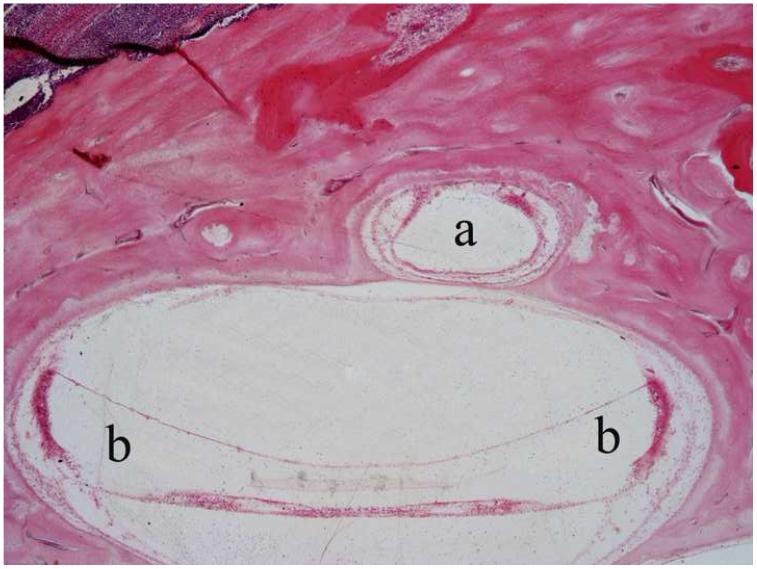
Objective: To demonstrate that what appears to be endolymphatic hydrops of the apical scala media is normal anatomy.
Study design: Computer-generated three-dimensional reconstruction of the cochlear apex and tabulation of the number of cases with arched Reissner's membranes (pseudohydrops) versus flat membranes.
Setting: Temporal bone laboratory consisting of 809 documented pairs of temporal bones.
Subjects and methods: Archival temporal bone sections from 107 bones (65 patients) were used to determine the percentage of arched (pseudohydrops) versus flat Reissner's membranes. Two bones, one of each membrane shape, were randomly selected for computer-generated three-dimensional reconstructions showing the cochlear apical anatomy.
Results: An arched Reissner's membrane was found in 48.6 percent of bones. In the cochlear apex, Reissner's membrane appears to be distended, simulating hydrops, due to its transition from a conical structure to a triangle bounded by the basilar membrane with the organ of Corti, the stria vascularis, and Reissner's membrane. Membrane findings were similar in both ears in 73.8 percent of the bilateral cases studied. There were no significant relationships between membrane type and clinical characteristics.
Conclusion: What appears to be endolymphatic hydrops of the cochlear apex is the transition area of the cochlear duct from a conical shape at the extreme apex to the triangular shape found in the rest of the cochlea. The appearance of distension is dependent upon the cochlear length and the level of the microscopic section.
Copyright (c) 2010 American Academy of Otolaryngology-Head and Neck Surgery Foundation. Published by Mosby, Inc. All rights reserved.
Figures


References
-
- Yamashita T, Schuknecht HF. Apical endolymphatic hydrops. Arch Otolaryngol. 1982;108:463–6. - PubMed
-
- Lau SK, Linthicum FH., Jr. Apparent apical endolymphatic hydrops: computer-aided three-dimensional reconstruction and histologic study of the apical turn of the cochlear duct. Am J Otol. 1993;14:79–81. - PubMed
-
- Ulehlová L, Voldrich L, Janisch R. Correlative study of sensory cell density and cochlear length in humans. Hear Res. 1987;28:149–51. - PubMed
-
- Kawano A, Seldon HL, Clark GM. Computer-aided three-dimensional reconstruction in human cochlear maps: measurement of the lengths of organ of Corti, outer wall, inner wall, and Rosenthal’s canal. Ann Otol Rhinol Laryngol. 1996;105:701–9. - PubMed
-
- Sato H, Sando I, Takahashi H. Sexual dimorphism and development of the human cochlea. Computer 3-D measurement. Acta Otolaryngol. 1991;111:1037–40. - PubMed
Publication types
MeSH terms
Grants and funding
LinkOut - more resources
Full Text Sources
Miscellaneous
