

EGFRvIII antibody-conjugated iron oxide nanoparticles for magnetic resonance imaging-guided convection-enhanced delivery and targeted therapy of glioblastoma
- PMID: 20647323
- PMCID: PMC2912981
- DOI: 10.1158/0008-5472.CAN-10-1022
EGFRvIII antibody-conjugated iron oxide nanoparticles for magnetic resonance imaging-guided convection-enhanced delivery and targeted therapy of glioblastoma
Abstract
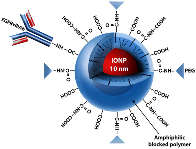
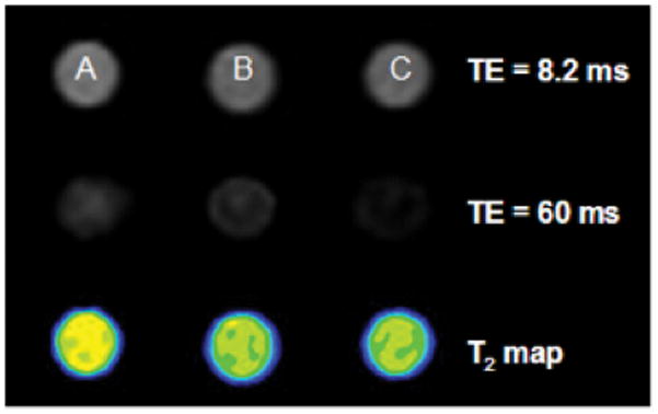
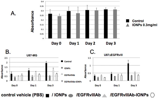
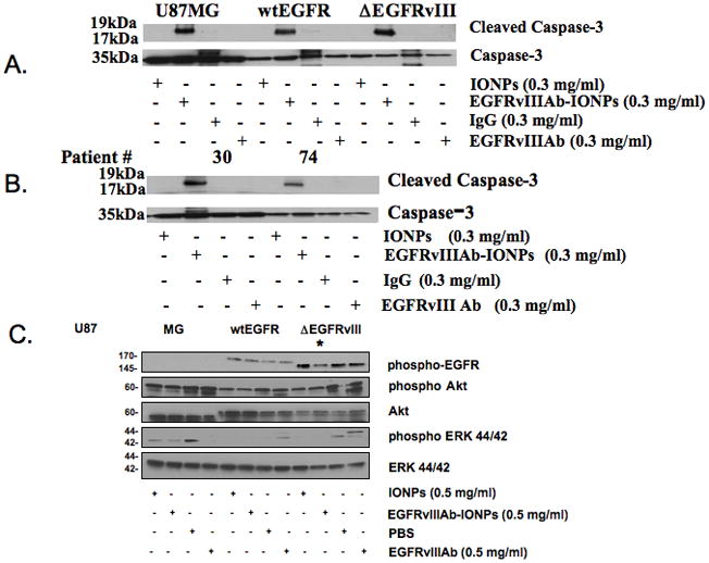
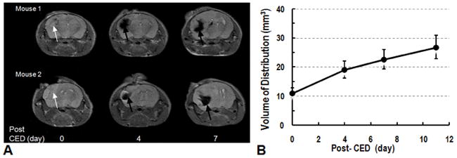
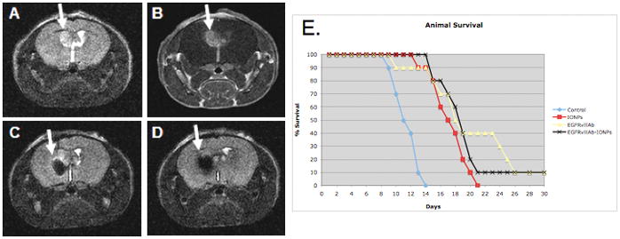
The magnetic nanoparticle has emerged as a potential multifunctional clinical tool that can provide cancer cell detection by magnetic resonance imaging (MRI) contrast enhancement as well as targeted cancer cell therapy. A major barrier in the use of nanotechnology for brain tumor applications is the difficulty in delivering nanoparticles to intracranial tumors. Iron oxide nanoparticles (IONP; 10 nm in core size) conjugated to a purified antibody that selectively binds to the epidermal growth factor receptor (EGFR) deletion mutant (EGFRvIII) present on human glioblastoma multiforme (GBM) cells were used for therapeutic targeting and MRI contrast enhancement of experimental glioblastoma, both in vitro and in vivo, after convection-enhanced delivery (CED). A significant decrease in glioblastoma cell survival was observed after nanoparticle treatment and no toxicity was observed with treatment of human astrocytes (P < 0.001). Lower EGFR phosphorylation was found in glioblastoma cells after EGFRvIIIAb-IONP treatment. Apoptosis was determined to be the mode of cell death after treatment of GBM cells and glioblastoma stem cell-containing neurospheres with EGFRvIIIAb-IONPs. MRI-guided CED of EGFRvIIIAb-IONPs allowed for the initial distribution of magnetic nanoparticles within or adjacent to intracranial human xenograft tumors and continued dispersion days later. A significant increase in animal survival was found after CED of magnetic nanoparticles (P < 0.01) in mice implanted with highly tumorigenic glioblastoma xenografts (U87DeltaEGFRvIII). IONPs conjugated to an antibody specific to the EGFRvIII deletion mutant constitutively expressed by human glioblastoma tumors can provide selective MRI contrast enhancement of tumor cells and targeted therapy of infiltrative glioblastoma cells after CED.
Figures
Similar articles
-
Radiosensitivity enhancement of radioresistant glioblastoma by epidermal growth factor receptor antibody-conjugated iron-oxide nanoparticles.J Neurooncol. 2015 Aug;124(1):13-22. doi: 10.1007/s11060-015-1807-0. Epub 2015 May 17. J Neurooncol. 2015. PMID: 25981803 Free PMC article.
-
Targeted therapy of glioblastoma stem-like cells and tumor non-stem cells using cetuximab-conjugated iron-oxide nanoparticles.Oncotarget. 2015 Apr 20;6(11):8788-806. doi: 10.18632/oncotarget.3554. Oncotarget. 2015. PMID: 25871395 Free PMC article.
-
Time-Resolved MRI Assessment of Convection-Enhanced Delivery by Targeted and Nontargeted Nanoparticles in a Human Glioblastoma Mouse Model.Cancer Res. 2019 Sep 15;79(18):4776-4786. doi: 10.1158/0008-5472.CAN-18-2998. Epub 2019 Jul 22. Cancer Res. 2019. PMID: 31331912 Free PMC article.
-
The evolution of the EGFRvIII (rindopepimut) immunotherapy for glioblastoma multiforme patients.Hum Vaccin Immunother. 2014;10(11):3322-31. doi: 10.4161/21645515.2014.983002. Hum Vaccin Immunother. 2014. PMID: 25625931 Free PMC article. Review.
-
An EGFRvIII-targeted bispecific T-cell engager overcomes limitations of the standard of care for glioblastoma.Expert Rev Clin Pharmacol. 2013 Jul;6(4):375-86. doi: 10.1586/17512433.2013.811806. Expert Rev Clin Pharmacol. 2013. PMID: 23927666 Free PMC article. Review.
Cited by
-
Clinical implications of microRNAs in human glioblastoma.Front Oncol. 2013 Feb 7;3:19. doi: 10.3389/fonc.2013.00019. eCollection 2013. Front Oncol. 2013. PMID: 23403472 Free PMC article.
-
Use of magnetic perfusion-weighted imaging to determine epidermal growth factor receptor variant III expression in glioblastoma.Neuro Oncol. 2012 May;14(5):613-23. doi: 10.1093/neuonc/nos073. Epub 2012 Apr 4. Neuro Oncol. 2012. PMID: 22492960 Free PMC article.
-
Cancer treatment by magneto-mechanical effect of particles, a review.Nanoscale Adv. 2020 Jun 19;2(9):3632-3655. doi: 10.1039/d0na00187b. eCollection 2020 Sep 16. Nanoscale Adv. 2020. PMID: 36132753 Free PMC article. Review.
-
An Insight to Brain Targeting Utilizing Polymeric Nanoparticles: Effective Treatment Modalities for Neurological Disorders and Brain Tumor.Front Bioeng Biotechnol. 2022 Feb 3;10:788128. doi: 10.3389/fbioe.2022.788128. eCollection 2022. Front Bioeng Biotechnol. 2022. PMID: 35186901 Free PMC article. Review.
-
The Emerging Applications of Nanotechnology in Neuroimaging: A Comprehensive Review.Front Bioeng Biotechnol. 2022 Jul 6;10:855195. doi: 10.3389/fbioe.2022.855195. eCollection 2022. Front Bioeng Biotechnol. 2022. PMID: 35875504 Free PMC article. Review.
References
-
- Singh SK, Hawkins C, Clarke ID, et al. Identification of human brain tumour initiating cells. Nature. 2004;432:396–401. - PubMed
-
- Li B, Yuan M, Kim IA, Chang CM, Bernhard EJ, Shu HK. Mutant epidermal growth factor receptor displays increased signaling through the phosphatidylinositol-3 kinase/AKT pathway and promotes radioresistance in cells of astrocytic origin. Oncogene. 2004;23:4594–602. - PubMed
Publication types
MeSH terms
Substances
Grants and funding
LinkOut - more resources
Full Text Sources
Other Literature Sources
Medical
Molecular Biology Databases
Research Materials
Miscellaneous
