. 2010 Sep 1;70(17):6725-34.
doi: 10.1158/0008-5472.CAN-10-0735. Epub 2010 Jul 20.

Tumor-specific CD8+ T cells expressing interleukin-12 eradicate established cancers in lymphodepleted hosts

Affiliations

Tumor-specific CD8+ T cells expressing interleukin-12 eradicate established cancers in lymphodepleted hosts

Sid P Kerkar et al. Cancer Res. .

Abstract

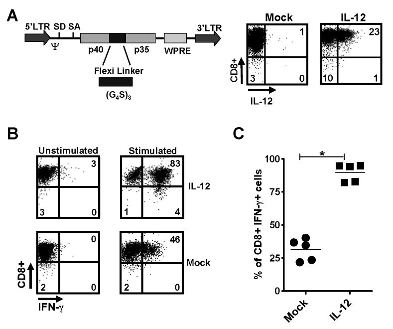
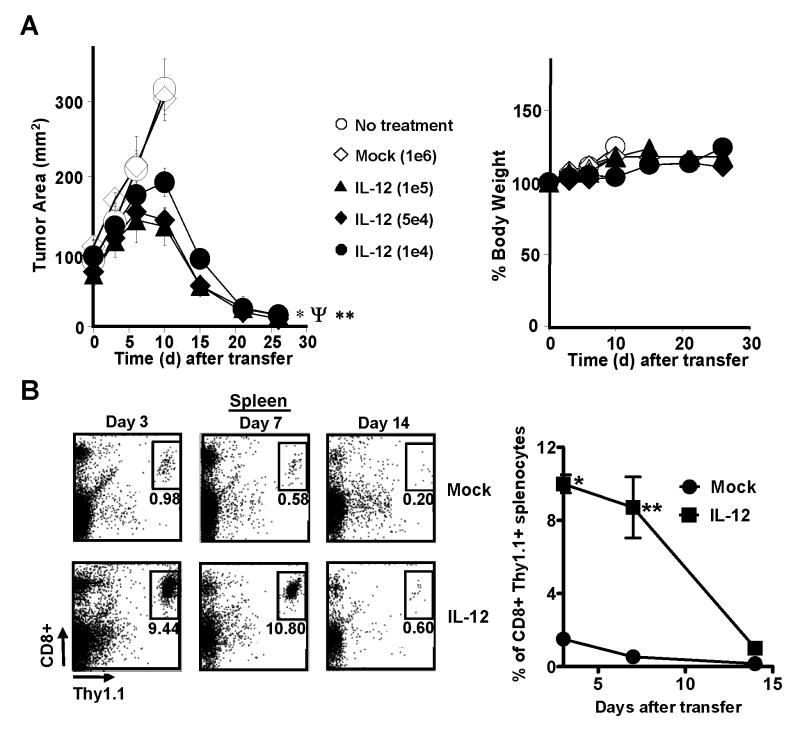
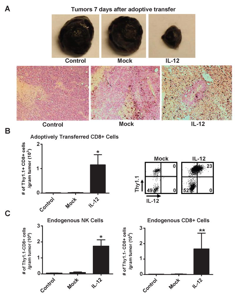
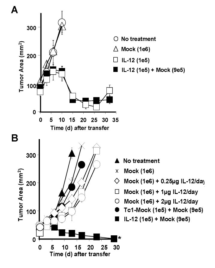
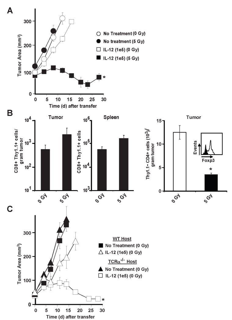
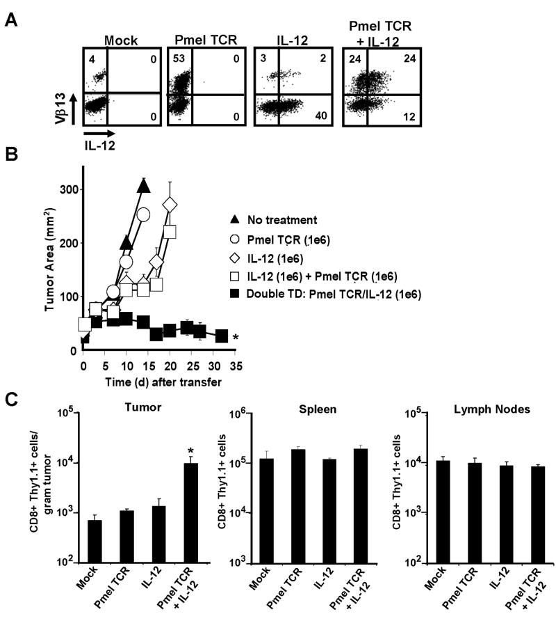
T-cell-based immunotherapies can be effective in the treatment of large vascularized tumors, but they rely on adoptive transfer of substantial numbers ( approximately 20 million) of tumor-specific T cells administered together with vaccination and high-dose interleukin (IL)-2. In this study, we report that approximately 10,000 T cells gene-engineered to express a single-chain IL-12 molecule can be therapeutically effective against established tumors in the absence of exogenous IL-2 and vaccine. Although IL-12-engineered cells did not perist long-term in hosts, they exhibited enhanced functionality and were detected in higher numbers intratumorally along with increased numbers of endogenous natural killer and CD8(+) T cells just before regression. Importantly, transferred T cells isolated from tumors stably overproduced supraphysiologic amounts of IL-12, and the therapeutic effect of IL-12 produced within the tumor microenvironment could not be mimicked with high doses of exogenously provided IL-12. Furthermore, antitumor effects could be recapitulated by engineering wild-type open-repertoire splenocytes to express both the single-chain IL-12 and a recombinant tumor-specific T-cell receptor (TCR), but only when individual cells expressed both the TCR and IL-12, indicating that arrested migration of T cells at the tumor site was required for their activities. Successful tumor eradication was dependent on a lymphodepleting preconditioning regimen that reduced the number of intratumoral CD4(+) Foxp3(+) T regulatory cells. Our findings reveal an approach to genetically modify T cells to reduce the cell number needed, eliminate the need for vaccines or systemic IL-2, and improve immunotherapy efficacy based on adoptive transfer of gene-engineered T cells.


Conflict of interest statement

The authors have no conflicting financial interests.

Figures

Figure 1
Figure 1
Pmel-1 CD8+ T cells engineered to overproduce IL-12 possess a distinct phenotype and enhanced functionality. A, The MSGV-1IL-12 retroviral vector (left panel), which encodes the p40 and p35 subunits of IL-12 linked by (Gly4Ser)3 flexible linker. SD, splice done; SA, splice accceptor; LTR, long terminal repeat; Ψ, packaging sequence. Pmel splenocytes were stimulated with 1 μM hgp10025–33 for 2 days, transduced with the MSGV-1IL-12 retrovirus, and analyzed by flow cytometry 3 days after for IL-12 expression (right panel). B, IL-12 transduced pmel-1 CD8+ cells from A were analyzed by intracellular staining without or with a 4hr secondary stimulation with phorbol 12-myristate 13-acetate (PMA) and ionomycin. C, The percentage of CD8+ IFN-γ+ cells following secondary stimulation in mock versus IL-12 transduced pmel cells was quantified (n=5; * p < 0.05). Experiments in A, B, and C are representative of at least 2 independent experiments.
Figure 2
Figure 2
Adoptive transfer of IL-12 engineered pmel-1 CD8+ T cells induces the regression of large, established tumors without exogenous IL-2 and vaccine. A, Anti-tumor activity following the adoptive transfer of 1×10⁵*, 5×10⁴Ψ, or 1×10⁴** pmel-1IL-12-TD cells into sublethaly-irradiated (5 Gy) mice bearing B16 tumors (n=5) established for 14 days (*, Ψ, **, p <0.05 compared no treatment) with no evidence of weight loss (right panel). B, 2.5×10⁵ mock versus IL-12 transduced pmel-1 thy1.1+ CD8+ T cells were transferred into sublethaly-irradiated (5 Gy) mice (n=3) and spleens were harvested on days 3, 7, and 14 after transfer and analyzed by flow cytometry for the percentage of transferred CD8+ thy1.1+ cells (left panel). B (right panel), Quantification of the percentage of CD8+ thy1.1+ cells in spleens (*,** p < 0.05 compared to mock). Experiments of A, and B are representative of at least 2 independent experiments.
Figure 3
Figure 3
Treatment with IL-12 engineered CD8+ T cells leads to increased tumor infiltration of adoptively transferred cells that stably expressing IL-12, and increased tumor infiltration by endogenous NK and CD8+ T cells. A, Representative subcutaneous tumor samples (top panel) excised 7 days (n=5) following the transfer of 5×10⁵ mock or IL-12 transduced pmel-1 CD8+ T cells into sublethaly-irradiated (5 Gy) mice bearing B16 tumors established for 14 days. Corresponding H and E stains (bottom panel; 100X magnification) for tumors in the top panel. B, 5×10⁵ mock versus IL-12 transduced pmel-1 thy1.1+ CD8+ T cells were transferred into sublethaly-irradiated (5Gy) mice (n=3), and tumors were excised 7 days after transfer, mechanically disrupted, and enumerated by flow cytometry for infiltration of adoptively transferred thy1.1+ CD8+ cells per gram of tumor (left panel; * p < 0.05 compared to control). B (right panel), IL-12 expression in tumor infiltrating thy1.1+ CD8+ cells (gated on the CD8+ population). C, Tumor samples from B, were also analyzed by flow cytometry for the number of endogenous thy1.1-, NK1.1+ cells (left panel; * p < 0.05 compared to control) and thy1.1-, CD8+ cells (right panel; ** p < 0.10 compared to control) per gram of tumor. All flow cytometry plots gated on live propidium iodide (PI)- populations. Experiments in A, B, and C representative of at least 2 independent experiments.
Figure 4
Figure 4
IL-12 engineered pmel-1 CD8+ T cells display enhanced anti-tumor responses compared to rIL-12 administered exogenously. A, Anti-tumor activity following the adoptive transfer of 1×10⁵ pmel-1IL-12-TD cells alone or in combination with 9×10⁵ mock transduced pmel-1 CD8+ T cells into sublethaly-irradiated (5Gy) mice bearing B16 tumors (n=5) established for 14 days. B, Treatment responses in sublethally-irradiated (5 Gy) tumor bearing mice (n=5) following the transfer of 1×10⁶ mock-transduced pmel-1 cells with exogenous rIL-12 daily administered intraperitoneally for 3 days (0.25 μg, 1 μg, or 2 μg), 1×10⁵ mock-transduced Tc1 cells (polarized with 3.33 ng ml-1 IL-12 ex vivo) or 1×10⁵ pmel-1IL-12-TD cells co-transferred with 9×10⁵ mock-transduced cells (* p<0.05 compared to all other treatment groups). Experiments in A and B are representative of at least 2 independent experiments.
Figure 5
Figure 5
Host irradiation (5 Gy) is required for anti-tumor immunity of adoptively transferred pmel-1IL-12-TD. A, Tumor treatment of sublethally-irradiated (5 Gy) or non-irradiated (0 Gy) WT B16 tumor bearing mice (n=5) treated with 1×10⁵ pmel-1IL-12-TD cells. B, Enumeration for thy1.1+ CD8+ T cells from isolated tumor samples and spleens (n=4) 6 days after the adoptive transfer of 5×10⁵ thy1.1+-marked pmel-1IL-12-TD into 0 Gy or 5 Gy treated hosts (left panel). Enumeration (n=5) for thy1.1- CD4+ T cells in tumor samples (right panel; * p<0.05 compared to 0 Gy treatment). Flow-cytometry of isolated thy1.1- CD4+ T cells for Foxp3 expression (right panel; gated on thy1.1-, CD4+ population). All flow cytometry samples gated on live propidium iodide (PI-)populations. C. Anti-tumor responses following the treatment of 1×10⁵ pmel-1IL-12-TD cells into non-ablated (0 Gy) WT or TCRα-/- B16 tumor bearing mice (n=5). Experiments in A and B are representative of at least 2 independent expermiments.
Figure 6
Figure 6
Tumor antigen-specific T-cell receptors are critical for the therapeutic responses of IL-12 engineered T cells. A, Cytofluorometric analysis of open-repertoire (WT) CD8+ T cells expressing IL-12 or pmel-1 TCR (Vβ-13+ staining) individually or on the same cell following retroviral transduction. All plots gated on CD8+ cells. B, Anti-tumor responses in sublethally-irradiated (5 Gy) tumor-bearing WT mice (n=5) treated with 1×10⁶ single-transduced cells (expressing either the pmel-1 TCR or IL-12), a combination of both single transduced populations (2×10⁶ cells), or 1×10⁶ CD8+ T cells co-expressing both the pmel-1 TCR and IL-12 (* p <0.05 compared to all other treatments). C, Enumeration of tumor, spleen and draining lymph nodes (n=5) for adoptively transferred open-repertoire CD8+ thy1.1+ T cells engineered to express IL-12 and/or the pmel-1 TCR as in panel (A). * p <0.05, compared to mock. All flow cytometry samples gated on live propidium iodide (PI-)populations. Experiments in A, B, and C are representative of at least 2 independent experiments.

References

    1. Breart B, Lemaitre F, Celli S, Bousso P. Two-photon imaging of intratumoral CD8+ T cell cytotoxic activity during adoptive T cell therapy in mice. J Clin Invest. 2008;118(4):1390–7. - PMC - PubMed
    1. Boissonnas A, Fetler L, Zeelenberg IS, Hugues S, Amigorena S. In vivo imaging of cytotoxic T cell infiltration and elimination of a solid tumor. J Exp Med. 2007;204(2):345–56. - PMC - PubMed
    1. Marigo I, Dolcetti L, Serafini P, Zanovello P, Bronte V. Tumor-induced tolerance and immune suppression by myeloid derived suppressor cells. Immunol Rev. 2008;222:162–79. - PubMed
    1. Hwu P, Du MX, Lapointe R, Do M, Taylor MW, Young HA. Indoleamine 2,3-dioxygenase production by human dendritic cells results in the inhibition of T cell proliferation. J Immunol. 2000;164(7):3596–9. - PubMed
    1. Srivastava MK, Sinha P, Clements VK, Rodriguez P, Ostrand-Rosenberg S. Myeloid-derived suppressor cells inhibit T-cell activation by depleting cystine and cysteine. Cancer Res. 70(1):68–77. - PMC - PubMed

Publication types

MeSH terms