. 2010 Nov;9(11):2438-59.
doi: 10.1074/mcp.M110.001859. Epub 2010 Jul 20.

Quantitative proteomic analysis of A549 cells infected with human respiratory syncytial virus

Affiliations

Quantitative proteomic analysis of A549 cells infected with human respiratory syncytial virus

Diane C Munday et al. Mol Cell Proteomics. 2010 Nov.

Abstract

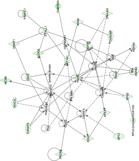
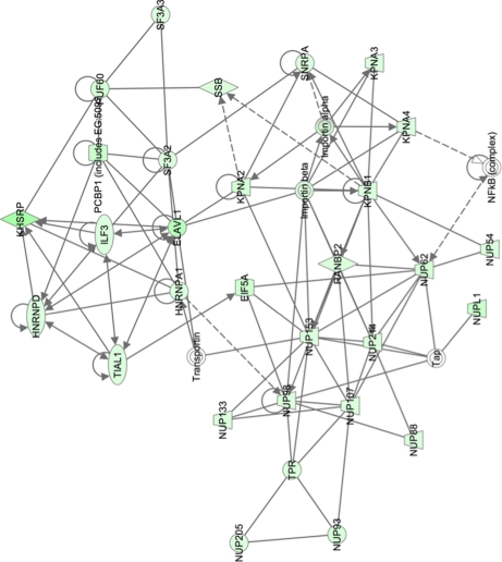
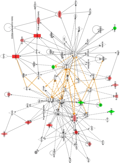
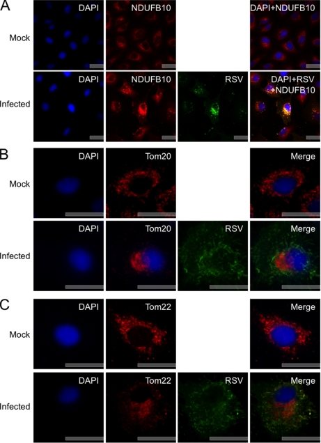
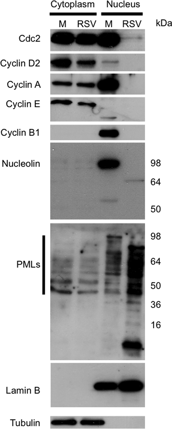
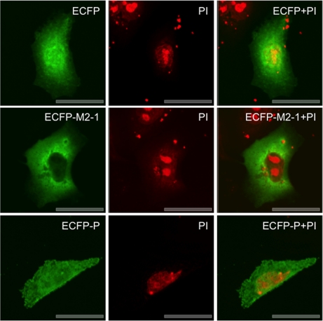
Human respiratory syncytial virus (HRSV) is a major cause of pediatric lower respiratory tract disease to which there is no vaccine or efficacious chemotherapeutic strategy. Although RNA synthesis and virus assembly occur in the cytoplasm, HRSV is known to induce nuclear responses in the host cell as replication alters global gene expression. Quantitative proteomics was used to take an unbiased overview of the protein changes in transformed human alveolar basal epithelial cells infected with HRSV. Underpinning this was the use of stable isotope labeling with amino acids in cell culture coupled to LC-MS/MS, which allowed the direct and simultaneous identification and quantification of both cellular and viral proteins. To reduce sample complexity and increase data return on potential protein localization, cells were fractionated into nuclear and cytoplasmic extracts. This resulted in the identification of 1,140 cellular proteins and six viral proteins. The proteomics data were analyzed using Ingenuity Pathways Analysis to identify defined canonical pathways and functional groupings. Selected data were validated using Western blot, direct and indirect immunofluorescence confocal microscopy, and functional assays. The study served to validate and expand upon known HRSV-host cell interactions, including those associated with the antiviral response and alterations in subnuclear structures such as the nucleolus and ND10 (promyelocytic leukemia bodies). In addition, novel changes were observed in mitochondrial proteins and functions, cell cycle regulatory molecules, nuclear pore complex proteins and nucleocytoplasmic trafficking proteins. These data shed light into how the cell is potentially altered to create conditions more favorable for infection. Additionally, the study highlights the application and advantage of stable isotope labeling with amino acids in cell culture coupled to LC-MS/MS for the analysis of virus-host interactions.


Figures

Fig. 1.
Fig. 1.
Quantitative proteomics using SILAC of HRSV-infected cells versus mock-infected cells. A, a diagrammatic representation of the methodology used in this analysis. Stable isotope (medium and heavy)-labeled amino acids were incorporated into newly synthesized cellular proteins. A549 cells were infected with HRSV (heavy) alongside a mock infection (medium), and 24 h postinfection subcellular fractionation was used to enrich the cytoplasmic and nuclear proteins. The infection efficiency and the fraction purity were then validated prior to sample preparation and LC-MS/MS analysis. B, indirect immunofluorescence confocal microscopy validation of HRSV infection efficiency in A549 cells 24 h postinfection. Staining with the HRSV antibody combination in mock-infected cells is shown as a control. Different magnifications are presented. Scale bars, 20 μm. C, Western blot confirmation of representative proteins enriched from the cytoplasmic and the nuclear fractions of mock-infected (M) and HRSV-infected (I) cells. Membrane-immobilized proteins were detected with tubulin (∼55 kDa) and lamin B1 (∼58 kDa) antibodies. Tubulin and lamin B1 were predominately localized in the cytoplasmic and nuclear fractions, respectively. D, Western blot confirmation of HRSV infection (and lack thereof in mock-infected cells). An anti-HRSV-specific polyclonal primary antibody was detected by an HRP conjugate. The locations of molecular mass markers (kDa) are indicated on the left, and the tentative assignments of proteins are indicated on the right. RSV, respiratory syncytial virus.
Fig. 2.
Fig. 2.
Classification of cellular proteins in HRSV-infected A549 cells according to their assigned fraction and biological function. For orientation, proteins classified as being involved in protein synthesis are to the right of the 12 o'clock position followed clockwise by protein synthesis, RNA post-transcriptional modification, cellular growth and proliferation, protein degradation (cytoplasmic fraction only), gene expression, and RNA trafficking. Formal descriptions of the different assigned functions are presented in supplemental Table 8.
Fig. 3.
Fig. 3.
Network pathway analysis of proteins identified in nuclear fraction that are primarily involved in cell growth and transcription regulation. Proteins shaded in green indicate a 2-fold or greater decrease in abundance in the nuclear fraction of HRSV-infected cells compared with mock-infected cells, and the color intensity corresponds to the degree of abundance. Proteins in white are those identified through the Ingenuity Pathways Knowledge Base. The shapes denote the molecular class of the protein. A solid line indicates a direct molecular interaction, and a dashed line indicates an indirect molecular interaction. A full explanation of lines and relationships is provided in supplemental Fig. 3.
Fig. 4.
Fig. 4.
Network pathway analysis of proteins identified in nuclear fraction that are primarily involved in molecular transport, including protein and RNA trafficking. Proteins shaded in green indicate a 2-fold or greater decrease in abundance in the nuclear fraction of HRSV-infected cells compared with mock-infected cells, and the color intensity corresponds to the degree of abundance. Proteins in white are those identified through the Ingenuity Pathways Knowledge Base. The shapes denote the molecular class of the protein. A solid line indicates a direct molecular interaction, and a dashed line indicates an indirect molecular interaction. A full explanation of lines and relationships is provided in supplemental Fig. 3.
Fig. 5.
Fig. 5.
Merged network pathway analysis of proteins involved in cellular assembly and organization, cellular compromise, and protein folding that were identified in cytoplasmic fraction. Proteins shaded in green indicate a 2-fold or greater decrease in abundance, and proteins shaded in red correspond to a 2-fold or greater increase in the cytoplasmic fraction of HRSV-infected cells. The color intensity denotes the degree of abundance. Proteins in white are those identified through the Ingenuity Pathways Knowledge Base. The shapes denote the molecular class of the protein. A solid line indicates a direct interaction, and a dashed line indicates an indirect interaction. The yellow lines denote the linkage between the pathways through NF-κB and STAT1 signaling. A full explanation of lines and relationships is provided in supplemental Fig. 3.
Fig. 6.
Fig. 6.
Alterations in mitochondrial protein abundance and mitochondrial integrity in HRSV-infected cells. A diagrammatic representation shows the inner and outer mitochondrial membranes with representative proteins identified in the quantitative proteomic analysis shown in their appropriate mitochondrial localizations. In the outer mitochondrial membrane, Tom20 and Tom22 interact with other Toms (some of which were identified in this analysis) to form a pore through which premitochondrial proteins are transported. Transmembrane pores such as VDACs may participate in the formation of the permeability transition pore complex, which is responsible for the release of mitochondrial products such as cytochrome c that trigger apoptosis. The inner membrane contains the protein complexes involved in the electron transport chain. Respiratory Complexes 1, 3, and 4 are proton pumps. Those proteins involved in cell proliferation and integrity such as PHB subunits PHB1 and PHB2 are also found in mitochondria.
Fig. 7.
Fig. 7.
A, indirect immunofluorescence confocal microscopy analysis of the subcellular localization of NDUFB10 in mock- and HRSV-infected A549 cells 24 h postinfection. NDUFB10 proteins are stained red, HRSV proteins are shown in green, and the nuclei are stained blue with DAPI. A merge image is also presented. B and C, indirect immunofluorescence confocal microscopy analysis of examples of outer mitochondrial membrane proteins whose abundance was shown to change in the quantitative proteomic analysis of the subcellular localization of Tom20 and Tom22 in mock- and HRSV-infected A549 cells 24 h postinfection. Tom20 (B) and Tom22 (C) proteins are stained red, HRSV proteins are green, and nuclei are stained blue with DAPI. A merge image is also presented. Scale bars, 20 μm. RSV, respiratory syncytial virus.
Fig. 8.
Fig. 8.
Indirect immunofluorescence confocal microscopy analysis of examples of mitochondrial transmembrane and inner membrane proteins whose abundance was shown to change in quantitative proteomic analysis of subcellular localization of VDAC1 and PHB in mock- and HRSV-infected A549 cells 24 h postinfection. VDAC1 proteins (A) and PHB proteins (B) are stained red, HRSV proteins are green, and nuclei are stained blue with DAPI. A merge image is also presented. Scale bars, 20 μm. RSV, respiratory syncytial virus.
Fig. 9.
Fig. 9.
Live cell confocal microscopy analysis of mitochondrial transition pore activity in mock- and HRSV-infected A549 cells 24 h postinfection. Nuclei are stained blue with Hoechst. All mitochondria are stained red with MitoTracker (A), healthy cells are also stained green with calcein AM (B), which is concentrated in the mitochondria. Merged images are also presented (C). Positive control cells were treated with ionomycin to allow the entry of excess calcium into the cells to trigger mitochondrial pore activation, which leads to the release of calcein AM and therefore the loss of green florescence. A large proportion of mitochondria in HRSV-infected cells were stained red and only weakly green, indicating that HRSV infection affected mitochondrial transition pore activity. Scale bars, 20 μm. Note that indirect immunofluorescence confocal microscopy was used to demonstrate that cells were infected with HRSV, and these data are presented in supplemental Fig. 2.
Fig. 10.
Fig. 10.
Potential disruption of proteins involved in nucleocytoplasmic trafficking in HRSV-infected cells. A, a diagrammatic representation of the nuclear pore complex showing examples of the nuclear pore proteins whose abundance was altered in the quantitative proteomic analysis of HRSV-infected versus mock-infected cells and also nucleocytoplasmic trafficking proteins. Many of the nups interact with one another and other nuclear envelope proteins; for example, nup53 interacts with nup98 and lamin B. B, indirect immunofluorescence confocal microscopy analysis of nuclear protein lamin B subcellular localization in mock- and HRSV-infected A549 cells 24 h postinfection. Lamin B proteins are stained red, HRSV proteins are shown in green, and the nuclei are stained blue with DAPI. A merge image is also presented. Scale bars, 20 μm. RSV, respiratory syncytial virus.
Fig. 11.
Fig. 11.
A, Western blot analysis of the cell cycle regulatory proteins and components of subnuclear structures in HRSV-infected (RSV) versus mock-infected cells (M) present in cytoplasmic and nuclear fractions. To validate fraction enrichment and loading control, tubulin and lamin B were used as markers for the cytoplasm and nucleus, respectively.
Fig. 12.
Fig. 12.
Indirect immunofluorescence confocal microscopy analysis of ND10s in HRSV-infected versus mock-infected cells at 24 h postinfection (A) and 36 h postinfection (B) is shown. PML proteins (the major constituent of ND10s) are stained red, the HRSV proteins are green, and the DNA in nuclei is stained blue with DAPI. A merge image is also presented. C, direct immunofluorescence confocal microscopy analysis of the subcellular localization of recombinant FLAG-tagged PML isoforms PML-I and PML-II in mock- and HRSV-infected A549 cells 24 h postinfection. PML-I and PML-II proteins are red, the HRSV proteins are green, and the nuclei are stained blue with DAPI. Scale bars, 20 μm. RSV, respiratory syncytial virus.
Fig. 13.
Fig. 13.
Direct immunofluorescence confocal microscopy analysis of overexpression analysis of ECFP, ECFP-M2-1, and ECFP-P in A549 cells 48 h post-transfection. ECFP is false colored green, and DNA in the nuclei is stained red with propidium iodide (PI).

References

    1. Hall C. B., Weinberg G. A., Iwane M. K., Blumkin A. K., Edwards K. M., Staat M. A., Auinger P., Griffin M. R., Poehling K. A., Erdman D., Grijalva C. G., Zhu Y., Szilagyi P. (2009) The burden of respiratory syncytial virus infection in young children. N. Engl. J. Med. 360, 588–598 - PMC - PubMed
    1. Openshaw P. J., Dean G. S., Culley F. J. (2003) Links between respiratory syncytial virus bronchiolitis and childhood asthma: clinical and research approaches. Pediatr. Infect. Dis. J. 22, S58–S64; discussion S64–S65 - PubMed
    1. Martinez F. D. (2003) Respiratory syncytial virus bronchiolitis and the pathogenesis of childhood asthma. Pediatr. Infect. Dis. J. 22, S76–82 - PubMed
    1. Collins P. L., Murphy B. R. (2002) Respiratory syncytial virus: reverse genetics and vaccine strategies. Virology 296, 204–211 - PubMed
    1. Collins P. L., Graham B. S. (2008) Viral and host factors in human respiratory syncytial virus pathogenesis. J. Virol. 82, 2040–2055 - PMC - PubMed

Publication types

MeSH terms

LinkOut - more resources