. 2010 Nov;38(20):6976-84.
doi: 10.1093/nar/gkq576. Epub 2010 Jul 6.

Repair of mitomycin C mono- and interstrand cross-linked DNA adducts by UvrABC: a new model

Affiliations

Repair of mitomycin C mono- and interstrand cross-linked DNA adducts by UvrABC: a new model

Mao-wen Weng et al. Nucleic Acids Res. 2010 Nov.

Abstract

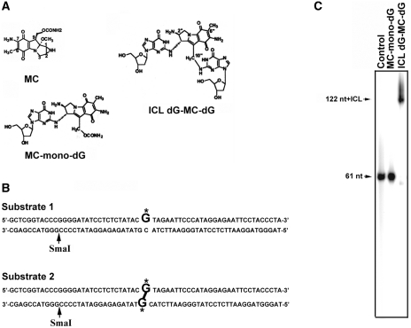
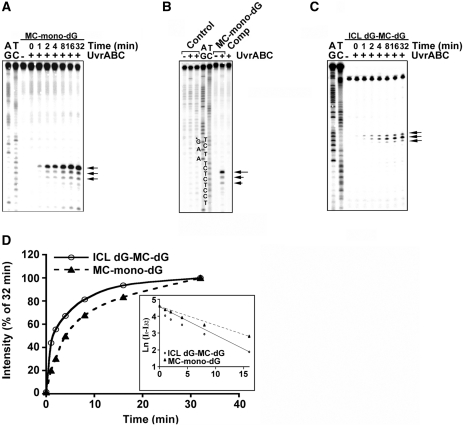
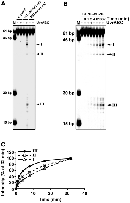
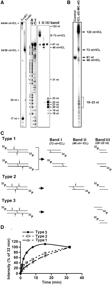
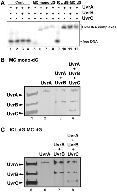
Mitomycin C induces both MC-mono-dG and cross-linked dG-adducts in vivo. Interstrand cross-linked (ICL) dG-MC-dG-DNA adducts can prevent strand separation. In Escherichia coli cells, UvrABC repairs ICL lesions that cause DNA bending. The mechanisms and consequences of NER of ICL dG-MC-dG lesions that do not induce DNA bending remain unclear. Using DNA fragments containing a MC-mono-dG or an ICL dG-MC-dG adduct, we found (i) UvrABC incises only at the strand containing MC-mono-dG adducts; (ii) UvrABC makes three types of incisions on an ICL dG-MC-dG adduct: type 1, a single 5' incision on 1 strand and a 3' incision on the other; type 2, dual incisions on 1 strand and a single incision on the other; and type 3, dual incisions on both strands; and (iii) the cutting kinetics of type 3 is significantly faster than type 1 and type 2, and all of 3 types of cutting result in producing DSB. We found that UvrA, UvrA+UvrB and UvrA+UvrB+UvrC bind to MC-modified DNA specifically, and we did not detect any UvrB- and UvrB+UvrC-DNA complexes. Our findings challenge the current UvrABC incision model. We propose that DSBs resulted from NER of ICL dG-MC-dG adducts contribute to MC antitumor activity and mutations.


Figures

Figure 1.
Figure 1.
(A) Chemical structures of MC, MC-mono-dG, and ICL dG-MC-dG DNA adducts. (B) The DNA sequence of the 61-bp DNA fragment containing a site-specific MC-mono-dG (substrate 1) or an ICL dG-MC-dG adduct (substrate 2). G* represents MC-modified deoxyguanosine. Restriction enzyme SmaI site is indicated. (C) Substrate 1, substrate 2 and control 61-bp DNA fragments were 5′-end 32P labeled, denatured and then separated by electrophoresis in a denaturing gel.
Figure 2.
Figure 2.
Kinetics of UvrABC incision of MC-mono-dG and ICL dG-MC-dG adducts. In (A) the MC-mono-dG–containing strand of substrate 1 was 5′-32P-end labeled. In (B) the MC-mono-dG–containing strand of substrate 1, the non-modified strand (Comp) of substrate 1, and control 61-bp fragments were 5′-32P-single-end labeled. In (C) substrate 2 was 32P-labeled at the 5′ end on both strands and digested with SmaI to generate single 5′-end-32P-labeled DNA fragments. The DNA fragments were incubated with UvrABC for different time periods (A and C), or for 60 min (B), and the resultant DNAs were separated by electrophoresis in a 12% polyacrylamide denaturing gel. (A–C) represent typical autoradiographs, and (D) represents the quantitations. The band intensities at different times (It) were normalized to the intensity at 32 min (I32), which encompasses 90% of total activity. The inset shows a semilogarithmic plot; the slopes yield two apparent first-order rate constants, Kobs,ABC, mono = 0.111 min−1 and Kobs,ABC, ICL = 0.169 min−1. Symbols: AG and TC represent Maxam and Gilbert sequencing reaction products (42); G*, MC-modified guanine; the arrows indicate the UvrABC incision bands; MC-mono-dG (filled triangle with dashed line); and ICL dG-MC-dG adducts (open circle with continous line).
Figure 3.
Figure 3.
Double-stranded DNA break formation resulted from UvrABC incision of an ICL dG-MC-dG adduct. (A) 32P-labeled substrate 1, substrate 2 and control 61-bp DNA fragments were reacted with UvrABC for 60 min and then separated in a nondenaturing polyacrylamide gel. (B) The kinetics of DSB formation resulted from UvrABC incision of an ICL dG-MC-dG DNA adduct. The 5′-end-32P-labeled 61-bp DNA fragments containing a site-specific ICL dG-MC-dG–DNA adduct were incubated with UvrABC nucleases as described in Figure 2. At different time periods of incubation, the resultant DNAs were separated in a nondenaturing polyacrylamide gel. The double-stranded DNA fragments standards are shown in lane M. (C) Quantitations. The band intensities at different times (It) shown in (B) were normalized to the intensity at 32 min (I32), which encompasses 90% of total activity. The data were plotted the same as in Figure 2.
Figure 4.
Figure 4.
Identification of the products resulting from UvrABC incision of the 61-bp DNA fragments containing an ICL dG-MC-dG–DNA adduct. (A) The three major bands (I, II and III) as shown in Figure 3 were extracted and separated by electrophoresis in a 12% polyacrylamide denaturing gel as described in Figure 2. Note: the DNA fragments in band I resulted in a band corresponding to size of ∼73 nt + ICL, the DNA fragments in band II resulted in a band corresponding size of ∼46 nt + ICL, and the DNA fragments in band III resulted a band of size ∼22-nt bands. The size standards were generated using the 61– bp DNA fragment containing an ICL dG-MC-dG adduct cut with EcoRI or EcoRV. The 61-nt band results from an unreacted 61-mer contamination during the strand construction. AG and TC represent Maxam and Gilbert sequencing reaction products (42). (B) 32P-labeled 61-bp DNA fragments containing an ICL dG-MC-dG DNA adduct were incubated with UvrABC for 32 min and the resultant DNAs were separated by electrophoresis the same as in (A). (C) The three possible types (1, 2 and 3) of UvrABC incision on the double-stranded 61-bp fragments containing a site-specific ICL dG-MC-dG DNA adduct that would result in generating fragments of the following approximate sizes: 73 nt + ICL, 46 nt + ICL and 22 nt. The arrows indicate the UvrABC cutting sites. For clarity, only the major incision positions are shown. It should be noted that type 1, 2 and 3 incisions resulted in producing DSB. (D) The kinetics of the three types of UvrABC cutting on the ICL dG-MC-dG lesion. The calculations were based on the band intensity shown in Figure 3B and the cutting models presented in Figure 4C.
Figure 5.
Figure 5.
The binding pattern of Uvr proteins with 32P-labeled DS 61-bp DNA fragments with and without a site-specific MC-mono-dG or an ICL dG-MC-dG adduct. In (A) the DNA fragments were incubated with UvrA, UvrA + UvrB and UvrA + UvrB + UvrC for 20 min and the resultant complexes were separated by electrophoresis in a 4.5% non-denaturing polyacrylamide gel containing 50 mM MgCl2 and 1 mM ATP at 2°C in 0.5 × TBE buffer for 2 h (lanes 1–12). The Uvr–DNA complexes are: (UvrA)2–DNA (lane 6); (UvrA)2-(UvrB)–DNA (lane 7); (UvrA)2-(UvrB)-(UvrC)–DNA (lane 8); (UvrA)4–DNA (lane 10); (UvrA)4-(UvrB)2–DNA (lane 11) and (UvrA)4-(UvrB)2-(UvrC)2–DNA (lane 12). To determine the Uvr protein composition in the Uvr–DNA complexes resulting from Uvr-bound DS 61-bp DNA fragments containing a site-specific (B) MC-mono-dG or (C) ICL dG-MC-dG adduct, bands isolated from Figure 5A were denatured by heat (95°C in 2% SDS for 5 min), separated in an 8% SDS-PAGE, and stained with silver stain. The Uvr proteins added to the DNA fragments that resulted in the different retarded bands are shown on the top of the panel. In (B) lane 2 is from lane 6 of Figure 5A; lane 3 is from lane 7 of Figure 5A; lane 4 is from lane 8 of Figure 5A In (C) lane 6 is from lane 10 of Figure 5A; lane 7 is from lane 11 of Figure 5A; lane 8 is from lane 12 of Figure 5A. Lanes 1 and 5 are the UvrA, UvrB and UvrC standards. Note: The complexes in each band contain all the added Uvr protein (i.e. UvrA was found in the MC-modified DNA added with UvrA; UvrA and UvrB were found in MC-modified DNA added with these two proteins; and UvrA, UvrB and UvrC were found in the MC-modified DNA added with these three proteins).

Similar articles

Cited by

References

    1. Kusenda Z, Kerger J, Awada A, Geurs F, Van Vreckem A, Habboubi N, Piccart MJ. Mitomycin C and vinblastine in combination with amifostine in metastatic breast cancer. A feasibility study of the EORTC–Investigational Drug Branch for Breast Cancer (IDBBC) Support Care Cancer. 1997;5:414–416. - PubMed
    1. Lenaz L. Mitomycin C in advanced breast cancer. Cancer Treat. Rev. 1985;12:235–249. - PubMed
    1. Bolenz C, Cao Y, Arancibia MF, Trojan L, Alken P, Michel MS. Intravesical mitomycin C for superficial transitional cell carcinoma. Expert Rev. Anticancer Ther. 2006;6:1273–1282. - PubMed
    1. Snegovoy A, Manzuk L, Artamonova E. Mitomycin plus capecitabine in metastatic breast cancer. 2008 Breast Cancer Symposium. 2008 General Poster Session E, Abstract No: 264.
    1. Bizanek R, McGuinness BF, Nakanishi K, Tomasz M. Isolation and structure of an intrastrand cross-link adduct of mitomycin C and DNA. Biochemistry. 1992;31:3084–3091. - PubMed

Publication types