Review
. 2010 Jul;3(4):537-46.
doi: 10.1161/CIRCHEARTFAILURE.109.903773.

Making the case for skeletal myopathy as the major limitation of exercise capacity in heart failure

Affiliations
Review

Making the case for skeletal myopathy as the major limitation of exercise capacity in heart failure

Holly R Middlekauff. Circ Heart Fail. 2010 Jul.
No abstract available


Figures

Figure 1
Figure 1
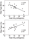
Panel A. Fiber type shift correlates with decreased exercise capacity. Morphometric analyses of muscle biopsies from HF patients have revealed a shift in fiber type, from slow twitch, oxidative type I fibers to fast twitch, glycolytic type IIb fibers. The shift in fiber type correlates with diminished exercise capacity as estimated by peak oxygen consumption. Panel B. Decreased mitochondrial density correlates with decreased exercise capacity. Quantified by electron microscopy, volume density of the mitochondrial in HF is significantly diminished compared to controls. See text for discussion. Filled squares=HF, open squares=controls. Reprinted with permission from reference .
Figure 1
Figure 1
Panel A. Fiber type shift correlates with decreased exercise capacity. Morphometric analyses of muscle biopsies from HF patients have revealed a shift in fiber type, from slow twitch, oxidative type I fibers to fast twitch, glycolytic type IIb fibers. The shift in fiber type correlates with diminished exercise capacity as estimated by peak oxygen consumption. Panel B. Decreased mitochondrial density correlates with decreased exercise capacity. Quantified by electron microscopy, volume density of the mitochondrial in HF is significantly diminished compared to controls. See text for discussion. Filled squares=HF, open squares=controls. Reprinted with permission from reference .
Figure 2
Figure 2
Correlation between exercise capacity and muscle oxidative capacity. HF patients have lower exercise capacity but similar muscle oxidative capacity compared to sedentary controls; both exercise capacity and muscle oxidative capacity are lower in these groups compared with active controls. The decreased exercise capacity in HF, therefore, cannot be entirely attributed to decreased muscle oxidative capacity. Open circles=HF, open triangles=sedentary controls, closed triangles=active controls. Modified from reference .
Figure 3
Figure 3
Abnormal excitation-contraction coupling in HF. See text for discussion. Modified from reference .
Figure 4
Figure 4
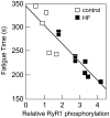
RyR1 phosphorylation and muscle fatigue. Time to fatigue was directly correlated to RyR1 PKA phosphorylation in rat skeletal muscle from sham operated (open squares) and HF (closed squares) animals. Reprinted with permission from reference .
Figure 5
Figure 5
Coordinated adaptation in oxygen transport. Arrow size indicates capacity; shaded arrows indicate sites of greatest limitation. The number of plus signs indicates the effect of changing capacity on changing maximal oxygen flux. The athlete has matched capacities at all steps. The untrained person is limited by cardiovascular delivery and muscle extraction. The chronic lung disease patient is limited by pulmonary diffusion. In chronic HF, the limited maximal cardiac output leads to down regulation of muscle oxygen extraction and mitochondrial utilization. Modified from reference .
Figure 6
Figure 6
Mechanisms by which sympathetic nerve activation may contribute to the skeletal myopathy of HF. See text for discussion.
Figure 7
Figure 7
Benefits of exercise. Exercise training decreases resting SNA, decreases inflammation, reverses morphometric and histochemical features of skeletal myopathy, and leads to improved blood flow, and increases quality of life and overall exercise capacity.
Figure 8
Figure 8
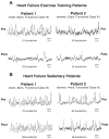
Sympathetic nerve recordings before and after exercise training period (panel A) or sedentary period (panel B). In pre-training patients and pre-sedentary control patients, SNA is markedly increased. Following the training period, but not the sedentary period, SNA is significantly reduced. Reproduced with permission from reference .

Similar articles

Cited by

References

    1. Vantura-Clapier R, De Sousa E, Veksler V. Metabolic myopathy in HF. New Physiol Sci. 2002;17:191–196. - PubMed
    1. Duscha B, Schulze PC, Robbins JL, Forman DE. Implications of chronic HF on peripheral vasculature and skeletal muscle before and after exercise training. Heart Fail Rev. 2008;13:21–37. - PubMed
    1. Troosters T, Gosselink R, Decramer M. COPD and chronic HF: Two muscle diseases? J Cardiopul Rehab. 2004;24:137–145. - PubMed
    1. Sinoway LI, Li J. A perspective on the muscle reflex:implications for congestive HF. J Appl Physiol. 2005;99:5–22. - PubMed
    1. Witte KK, Clark AL. Why does chronic HF cause breathlessness and fatigue? Prog Cardiovasc Dis. 2007;49:366–384. - PubMed

Publication types

MeSH terms