Review
. 2010 Sep;83(993):791-803.
doi: 10.1259/bjr/76002141. Epub 2010 Jul 20.

CT and MRI in the evaluation of extraspinal sciatica

Affiliations
Review

CT and MRI in the evaluation of extraspinal sciatica

T Ergun et al. Br J Radiol. 2010 Sep.

Abstract

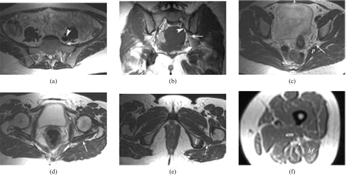
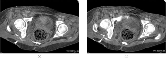
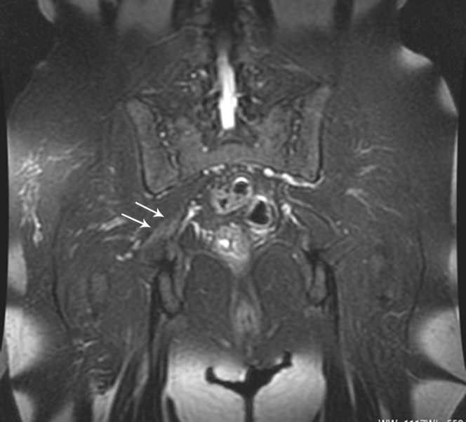
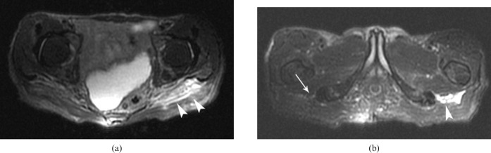
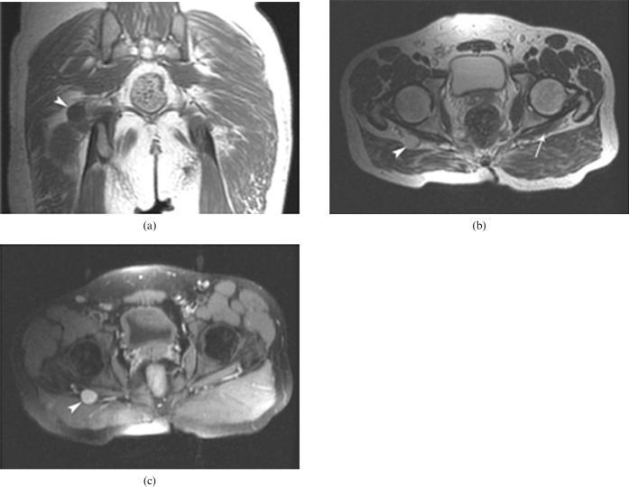
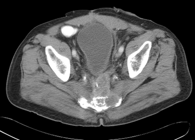
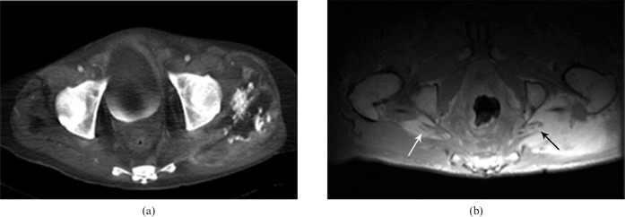
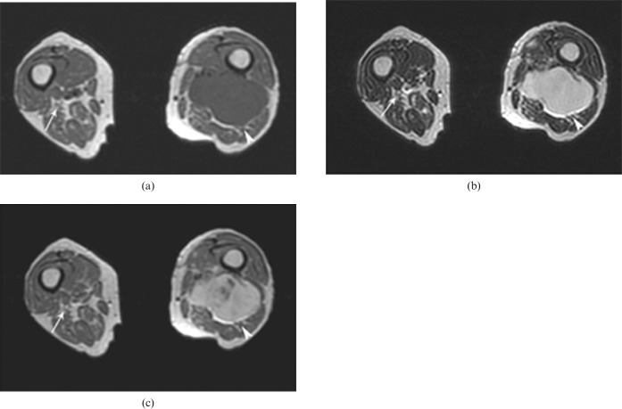
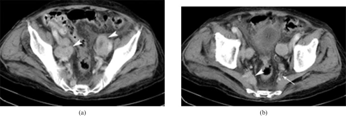
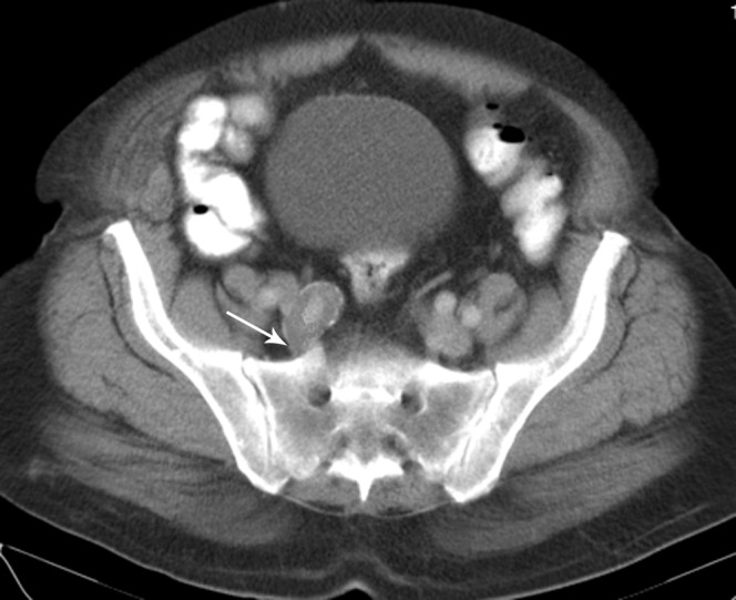
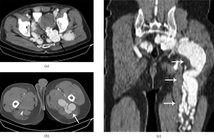
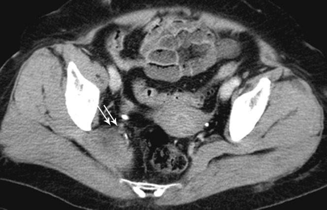
Sciatica is the most frequently encountered symptom in neurosurgical practice and is observed in 40% of adults at some point in their lives. It is described as pain of the hip and the lower extremity secondary to pathologies affecting the sciatic nerve within its intraspinal or extraspinal course. The most frequent cause is a herniating lumbar disc pressing on the neural roots. Extraspinal causes of sciatic pain are usually overlooked because they are extremely rare and due to intraspinal causes (lumbar spinal stenosis, facet joint osteoarthritis, fracture, and tumors of the spinal cord and spinal column) being the main consideration. Early diagnosis of sciatica significantly improves the likelihood of relieving symptoms, as well as avoiding any additional neurologic injury and unnecessary surgery. We evaluate histolopathologically confirmed extraspinal causes of sciatica cases, accompanied by their presented computed tomography and/or magnetic resonance imaging findings.


Figures

Figure 1
Figure 1
Normal MRI appearance of the lumbosacral trunk, sacral plexus and sciatic nerve. Axial T1 weighted image (a) at the level of the sacral wings shows the lumbosacral trunk (arrow) anterior to the sacrum and posterior to the iliac vessels (arrowhead). Coronal T1 weighted image (b) shows the normal sacral plexus (arrowhead) passing anterior to the sacral wings and continuing inferolaterally to leave the pelvis through the greater sciatic foramen (thin arrow) as the sciatic nerve (thick arrow). Axial T2 weighted image (c) at the level of the greater sciatic foramen shows the sacral plexus (arrow) anterior to the piriformis (P) muscle and lateral to the inferior gluteal vessels. Axial T2 weighted image (d) at the level of the gluteal region shows the sciatic nerve (arrow) superficial to the lateral rotator muscles and deep to the gluteus maximus muscle. Axial T2 weighted image (e) at the level of the infragluteal region shows the sciatic nerve (arrow) between the ischial tuberosity (i) and the greater trochanter. Axial T1 weighted image (f) at the mid-femoral level shows the sciatic nerve (arrow) between the adductor magnus (am) and biceps femoris (bf) muscles.
Figure 2
Figure 2
Post-traumatic heterotropic ossification in a 35-year-old female patient with a history of trauma sustained 6 years previously, and complaining of persistent numbness and pain in the right lower limb. Axial CT images (a,b) show a gluteus-level heterotropic ossification (with a lucent central portion) surrounding the right sciatic nerve (arrowhead). The left sciatic nerve appears normal (arrow).
Figure 3
Figure 3
Post-traumatic oedema in a 54-year-old female patient complaining of right foot numbness and pain radiating to the back of her right thigh following trauma. Coronal fat suppression T2 weighted image shows a right lumbosacral plexus oedema at the level of the greater sciatic foramen (arrows). The left lumbosacral plexus appears normal.
Figure 4
Figure 4
Footdrop in a 45-year-old female patient with an intramuscular injection history 3 days previously; axial fat suppression T2 weighted images (a,b) reveal oedema in the course of the sciatic nerve (arrowheads) The right sciatic nerve appears normal (arrow).
Figure 5
Figure 5
Sacral fracture in a 52-year-old male patient with a post-traumatic right footdrop. Axial CT image reveals a separation fracture affecting the neural foramina in the right half of the sacrum.
Figure 6
Figure 6
Intramuscular haematoma in a 51-year-old female patient with left-side sciatica, following a penetrating trauma. Axial T1 weighted (a) and T2 weighted (b) images show a large haematoma at the level of the greater sciatic foramen involving the left piriformis muscle, lumbosacral plexus and sciatic nerve (arrowheads). The right sciatic nerve appears normal (arrows).
Figure 7
Figure 7
Presacral abscess in a 55-year-old male patient with a left lumbosacral plexopath. A contrast-enhancing signal increase consistent with sacral osteomyelitis is observed on the fat suppression axial T1 weighted images following intravenous Gd administration (a) and on the axial fat suppression T2 weighted images (b). In addition, a presacral region cystic mass affecting the left lumbosacral plexus and consistent with an abscess is seen, demonstrating peripheral contrast attenuation (arrows). The right lumbosacral plexus appears normal (arrowhead) on the coronal T1 weighted images (c). The presacral abscess effect on the left lumbosacral plexus is clearly observable (arrows).
Figure 8
Figure 8
Sacroiliitis in a 19-year-old male patient with right-side sciatica. Coronal T1 weighted (a) and T2 weighted fat suppression (b) and axial T2 weighted fat suppression (c,d) MR images reveal sacroiliitis of the right sacroiliac joint and neighbouring oedema pressing on the lumbosacral plexus (arrows).
Figure 9
Figure 9
Schwannoma in a 37-year-old female patient complaining of right radicular leg pain. Coronal T1 weighted (a), axial T2 weighted images (b) and fat suppression T1 weighted images (c) following intravenous contrast administration reveals a smooth-contoured solid mass with significant contrast enhancement in the right sciatic nerve (arrowheads). The left sciatic nerve appears normal (arrow).
Figure 10
Figure 10
Colorectal carcinoma in a 64-year-old female patient with lumbosacral plexopathy. Axial CT image demonstrating a rectal carcinoma invading the left half of sacrum, the left S3 neural root and the left piriformis muscle.
Figure 11
Figure 11
Intramuscular metastasis in a 52-year-old male patient with a history of advanced stage gastric carcinoma complaining of waist pain, bilateral thigh pain (more severe on the left thigh), radiating to both legs, numbness and left leg cramps. (a) Pelvic CT image showing a solid left gluteal mass containing diffuse necrosis and calcification and peripheral contrast attenuation. (b) Axial fat suppression contrast-enhanced T1 weighted MR image demonstrating a well-demarcated solid mass in the left gluteal and right piriformis muscles with significant contrast enhancement. In addition, the sciatic nerves fibres are seen to be surrounded by tumour masses and appear thickened with no contrast enhancement (arrows).
Figure 12
Figure 12
Soft-tissue sarcoma in a 39-year-old female patient with left-side sciatica. Axial T1 weighted (a), T2 weighted (b) and T1 weighted images following intravenous contrast administration (c) demonstrating a soft-tissue mass with significant contrast enhancement in the posterior femoral compartment; the close relation of the mass to the sciatic nerve is seen (arrowheads). The right sciatic nerve appears normal (arrows).
Figure 13
Figure 13
Malignant osseous tumour in a 57-year-old male patient complaining of persistent left leg pain and numbness in the left foot. Axial T2 weighted (a), fat suppression contrast-enhanced T1 weighted (b) and fat suppression T2 weighted (c) MR images demonstrate a solid mass causing left acetabular destruction and have a soft-tissue component surrounding the infragluteal portion of the sciatic nerve (arrows). The right sciatic nerve appears normal (arrowhead).
Figure 14
Figure 14
Intramuscular lipoma in a 64-year-old male patient complaining of left foot numbness. Axial CT image shows a lipoma within the left external obturator muscle pressing on the left sciatic nerve (arrow). The right sciatic nerve appears normal (arrowhead).
Figure 15
Figure 15
Osteochondroma in a 35-year-old female patient complaining of right foot numbness. Axial T1 weighted images (a,b) showing a pedunculated osteochondroma located in the posterior portion of the femur and pressing on the sciatic nerve (arrowheads). The left sciatic nerve appears normal (arrows).
Figure 16
Figure 16
Ganglion cyst in a 51-year-old female patient with left-side sciatica; Axial T2 weighted (a) and fat-suppression T2 weighted (b) MR images demonstrating a round hyperintense mass along the course of the sciatic nerve at the posterior femoral compartment (arrow).
Figure 17
Figure 17
Lymphoma in a 35-year-old female patient with right buttock pain and normal lumbar MRI. Axial CT images (a) reveal multiple bilateral parailiac lymphadenopathies (arrowheads). At the level of the lower sacrum, CT (b) reveals enlarged lymph nodes pressing on the sciatic nerve in the vicinity of the right piriformis muscle (arrowhead). The left lumbosacral plexus appears normal (arrow).
Figure 18
Figure 18
A right internal iliac artery aneurysm in a 43-year-old male patient with lower back pain radiating to the right leg. Axial CT image demonstrating a partially thrombosed aneurysmal dilation of the right iliac artery pressing on the right lumbosacral plexus (arrow).
Figure 19
Figure 19
Arteriovenous fistula in a 49-year-old male patient with left sciatica and a history of penetrating trauma 2 years previously. Axial (a,b) and coronal reformatted (c) CT images demonstrating a thrombosed aneurysm (black arrow) and diffuse varicose veins (white arrows) related to the arteriovenous fistula pressing on the left lumbosacral plexus and sciatic nerve. The right sciatic nerve appears normal (arrowhead).
Figure 20
Figure 20
Piriformis syndrome in a 35-year-old female patient with a history of a traffic accident involvement 7 days previously, and complaining of persistent hip pain radiating to the posterior of her right thigh, as well as numbness in the right foot. Axial CT image reveals an intramuscular piriformis haematoma causing enlargement of the muscle. In addition, thickening and anterior displacement of the right sciatic nerve are seen (arrows).

Similar articles

Cited by

References

    1. Pick T, Howden R. Anatomy, descriptive and surgical (Gray's anatomy). Philadelphia, PA: Running Press, 1974: 781–93
    1. Moore KL, Dalley AF. Clinically oriented anatomy. Philadelphia, PA: Lippincott, Williams & Wilkins, 1999
    1. Maravilla KR, Bowen BC. Imaging of the peripheral nervous system: evaluation of peripheral neuropathy and plexopathy. AJNR Am J Neuroradiol 1998;19:1011–23 - PMC - PubMed
    1. Villarejo FJ, Pascual AM. Injection injury of the sciatic nerve (370 cases). Childs Nerv Syst 1993;9:229–32 - PubMed
    1. Ergun T, Lakadamyali H, Derincek A, Cagla Tarhan N, Ozturk A. Magnetic resonance imaging in the visualization of benign tumors and tumor-like lesions of hand and wrist. Curr Probl Diagn Radiol 2010;39:1–16 - PubMed