. 2010 Sep;30(18):4404-14.
doi: 10.1128/MCB.00358-10. Epub 2010 Jul 20.

Las1L is a nucleolar protein required for cell proliferation and ribosome biogenesis

Affiliations

Las1L is a nucleolar protein required for cell proliferation and ribosome biogenesis

Christopher D Castle et al. Mol Cell Biol. 2010 Sep.

Abstract

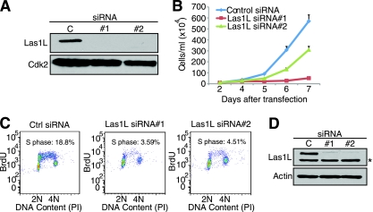
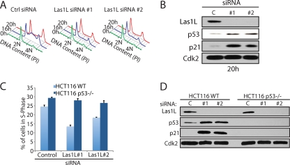
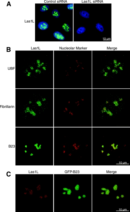
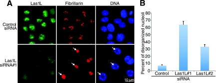
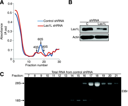
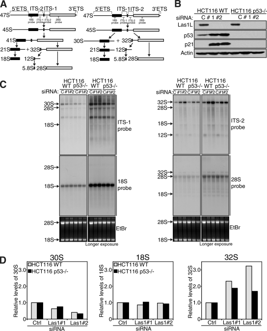
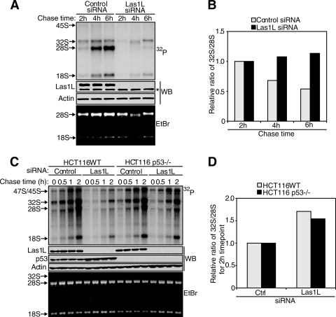
Ribosome biogenesis is a highly regulated process ensuring that cell growth (increase in biomass) is coordinated with cell proliferation. The formation of eukaryotic ribosomes is a multistep process initiated by the transcription and processing of rRNA in the nucleolus. Concomitant with this, several preribosomal particles, which transiently associate with numerous nonribosomal factors before mature 60S and 40S subunits are formed and exported in the cytoplasm, are generated. Here we identify Las1L as a previously uncharacterized nucleolar protein required for ribosome biogenesis. Depletion of Las1L causes inhibition of cell proliferation characterized by a G1 arrest dependent on the tumor suppressor p53. Moreover, we demonstrate that Las1L is crucial for ribosome biogenesis and that depletion of Las1L leads to inhibition of rRNA processing and failure to synthesize the mature 28S rRNA. Taken together, our data demonstrate that Las1L is essential for cell proliferation and biogenesis of the 60S ribosomal subunit.


Figures

FIG. 1.
FIG. 1.
Las1L protein is conserved throughout evolution. (A) Diagram of human Las1L protein, with the conserved Las1L domain and a C-terminal glutamic acid-rich region indicated. (B) Sequence alignment of human Las1L domain (residues 42 to 188) across 11 model organisms. Black shading denotes identical residues and conserved substitutions in 100% of the sequences, while gray shading indicates conservation in more than 50% of the sequences. Numbers in parentheses represent the gap lengths between residues. Organisms and GenBank sequence accession numbers are as follows: Homo sapiens, Q9Y4W2.2; Canis lupus familiaris, XP_538062.2; Bos taurus, NP_001070283.2; Mus musculus, A2BE28.1; Gallus gallus, Q5ZIQ0; Danio rerio, NP_001119872.1; Caenorhabditis elegans, Q9U1T1; Schizosaccharomyces pombe, O42936.1; Arabidopsis thaliana, Q8RXE3; Saccharomyces cerevisiae, P36146.1; and Drosophila melanogaster, Q8SZY9. Alignment was performed using ClustalX 2.0.12.
FIG. 2.
FIG. 2.
Las1L is required for cell proliferation. (A) HCT116 cells were transfected with nontargeting control (represented by the letter C), Las1L#1, or Las1L#2 siRNA. Depletion of Las1L at day 7 posttransfection was confirmed by Western blotting with a Las1-specific antibody. A Cdk2 antibody was used as a loading control. (B) Growth curve of HCT116 cells transfected with control siRNA or Las1L#1 and Las1L#2 siRNA as indicated. Twenty-four hours after transfection, cells were replated and counted at days 2, 4, 5, 6, and 7 posttransfection. Error bars indicate standard deviations. (C) BrdU and PI labeling of HCT116 cells transfected with control (Ctrl) or Las1L siRNAs. Seventy-two hours after transfection, the cells were analyzed by flow cytometry for DNA content. The percentage of cells in S phase is indicated on each graph. (D) Western blot analysis from protein lysates of cells used for the experiment with results shown in panel C, using anti-Las1L and antiactin antibodies to confirm knockdown. The asterisk indicates the presence of an unspecific band.
FIG. 3.
FIG. 3.
Depletion of Las1L induces a p53-dependent G1 cell cycle arrest. (A) Cell cycle profiles of HCT116 cells transfected with control (Ctrl), Las1L#1, or Las1L#2 siRNA. Twenty-four hours after transfection, cells were incubated in serum-free medium for an additional 72 h to synchronize the cells in G1. Serum was reintroduced, and the cell cycle phase was monitored at the indicated time points by staining with propidium iodide (PI) followed by analysis by flow cytometry. (B) Western blot analysis of cells from the 20-h time point release described for panel A, with specific antibodies against Las1L, p53, p21, and Cdk2. (C) Flow cytometry analysis of HCT116 WT and p53-null cells transfected with control, Las1L#1, or Las1L#2 siRNA. Seventy-two hours after transfection, cells were analyzed for BrdU incorporation, and the results are graphed as percentages of cells in S phase. Data represent the means from 3 independent experiments. Error bars indicate standard deviations. (D) Western blot analysis from protein lysates of the cells used for the experiments with results shown in panel C, with anti-Las1L, anti-p53, anti-p21, and anti-Cdk2 antibodies.
FIG. 4.
FIG. 4.
Las1L localizes to the nucleolus. (A) Immunofluorescence analysis of U2OS cells transfected with control or Las1L#1 siRNA. Seventy-two hours after transfection, cells were fixed and immunostained with anti-Las1L (green). DNA was visualized by staining with Hoechst 33342 (blue). The scale bar applies to both panels. (B) Confocal microscopy representing colocalization of Las1L (green) with nucleolar compartmental markers UBF, fibrillarin, and B23 (red) by immunostaining. Before staining, cells were fixed with 4% formaldehyde, except for B23, where methanol-acetone was used. The scale bar applies to all panels. (C) Colocalization of Las1L with B23 was confirmed using a GFP-B23 construct (green) and an anti-Las1L antibody (red). Cells were fixed with 4% formaldehyde. The scale bar applies to all panels.
FIG. 5.
FIG. 5.
Las1L depletion causes nucleolar disorganization. (A) Nucleolar disorganization in the absence of Las1L was analyzed by immunofluorescence. HCT116 cells were transfected with control or Las1L#1 siRNA for 72 h. Cells were fixed and immunostained with anti-Las1L antibody (green) or antifibrillarin antibody (red), and DNA was visualized with Hoechst 33342 (blue). The scale bar applies to all panels. Arrows indicate perturbed nucleoli. (B) The numbers of cells with disorganized nucleoli were counted in 5 different low-power fields and graphed. Error bars indicate standard deviations.
FIG. 6.
FIG. 6.
Depletion of Las1L causes defects in ribosome biogenesis. (A) Polysome profile of HCT116 cells infected with lentivirus-expressing control shRNA or Las1L shRNA 5 days after puromycin selection. Polysome profiles were performed by fractionation on a 10 to 50% sucrose gradient. Fractions were collected, and the optical density at 260 nm (A260) was measured. The positions of the 40S and 60S native subunits and the 80S monosomes are indicated. (B) Western blot analysis showing the levels of Las1L in control shRNA- or Las1L shRNA-infected cells used for the experiments with results shown in panel A. Actin levels were measured to the control level for protein loading. (C) RNA (stained with ethidium bromide [EtBr]) from fractions 7 to 21 was extracted and separated on a 1.2% formaldehyde gel to confirm the presence of the 40S or 60S subunit by monitoring the appearance of the 18S or 28S rRNA, respectively.
FIG. 7.
FIG. 7.
Las1L is required for proper processing of ITS-2. (A) Schematic representation (21) of the 47S rRNA primary transcript and the two major processing pathways, with indicated processed rRNA intermediates. The positions of the specific probes used for Northern blot analysis are indicated. (B) Western blot analysis with anti-Las1L, anti-p53, anti-p21, and antiactin antibodies to confirm knockdown of Las1L in the cells used for Northern blot analysis with results shown in panel C. (C) Northern blot analysis of total RNA from HCT116 WT or HCT116 p53−/− cells transfected with control (represented by the letter C), Las1L#1, or Las1L#2 siRNA for 72 h. Equal amounts of total RNA were hybridized with probes specific for ITS-1, ITS-2, 28S, and 18S rRNA intermediates, as indicated to the right of each panel. Right panels for each probe represent longer exposure times. (D) Quantification of relative band intensities of 32S, 30S, and 18S rRNAs in the shorter exposures shown in panel C. Intensities were normalized to the control level for each respective cell type for each panel.
FIG. 8.
FIG. 8.
Las1L is necessary for 28S rRNA synthesis. (A) Pulse-chase analysis of rRNA in HCT116 WT cells transfected with control or Las1L#1 siRNA. Cells were pulse-labeled with 32P-orthophosphate for 2 h and chased in normal growth medium for the indicated amounts of time. Total RNA was harvested and separated on a 1.2% formaldehyde denaturing gel. Knockdown of Las1L was verified by Western blot analysis (WB) with an anti-Las1L antibody. The asterisk indicates the presence of an unspecific band. Equal protein loading was confirmed using an antiactin antibody. The bottom panel represents an ethidium bromide (EtBr) RNA gel before transfer. (B) Quantification of band intensities from the experiment with results shown in panel A. 32S/28S ratios for the indicated chase times are shown. Intensities were normalized to the 32S/28S ratio at the 2-h time point. (C) Pulse-chase analysis of rRNA in HCT116 WT and HCT116 p53−/− cells. Cells were labeled as described for panel A and chased in normal growth medium for the indicated amounts of time. Knockdown of Las1L was verified by Western blot analysis with an anti-Las1 antibody. Levels of p53 were determined using an anti-p53 antibody. Equal protein loading was confirmed using an antiactin antibody. (D) Quantification of band intensities from the experiment with results shown in panel C. The relative 32S/28S ratio at the 2-h time point is shown. Intensities were normalized to the control level for each respective cell type.

Similar articles

Cited by

References

    1. Amsterdam, A., K. C. Sadler, K. Lai, S. Farrington, R. T. Bronson, J. A. Lees, and N. Hopkins. 2004. Many ribosomal protein genes are cancer genes in zebrafish. PLoS Biol. 2:E139. - PMC - PubMed
    1. Arabi, A., S. Wu, K. Ridderstrale, H. Bierhoff, C. Shiue, K. Fatyol, S. Fahlen, P. Hydbring, O. Soderberg, I. Grummt, L. G. Larsson, and A. P. Wright. 2005. c-Myc associates with ribosomal DNA and activates RNA polymerase I transcription. Nat. Cell Biol. 7:303-310. - PubMed
    1. Belin, S., A. Beghin, E. Solano-Gonzalez, L. Bezin, S. Brunet-Manquat, J. Textoris, A. C. Prats, H. C. Mertani, C. Dumontet, and J. J. Diaz. 2009. Dysregulation of ribosome biogenesis and translational capacity is associated with tumor progression of human breast cancer cells. PLoS One 4:e7147. - PMC - PubMed
    1. Bhat, K. P., K. Itahana, A. Jin, and Y. Zhang. 2004. Essential role of ribosomal protein L11 in mediating growth inhibition-induced p53 activation. EMBO J. 23:2402-2412. - PMC - PubMed
    1. Boisvert, F. M., S. van Koningsbruggen, J. Navascues, and A. I. Lamond. 2007. The multifunctional nucleolus. Nat. Rev. Mol. Cell Biol. 8:574-585. - PubMed