. 2010 Sep;30(18):4391-403.
doi: 10.1128/MCB.00002-10. Epub 2010 Jul 20.

The expression profile of the major mouse SPO11 isoforms indicates that SPO11beta introduces double strand breaks and suggests that SPO11alpha has an additional role in prophase in both spermatocytes and oocytes

Affiliations

The expression profile of the major mouse SPO11 isoforms indicates that SPO11beta introduces double strand breaks and suggests that SPO11alpha has an additional role in prophase in both spermatocytes and oocytes

Marina A Bellani et al. Mol Cell Biol. 2010 Sep.

Abstract

Both in mice and humans, two major SPO11 isoforms are generated by alternative splicing: SPO11alpha (exon 2 skipped) and SPO11beta. Thus, the alternative splicing event must have emerged before the mouse and human lineages diverged and was maintained during 90 million years of evolution, arguing for an essential role for both isoforms. Here we demonstrate that developmental regulation of alternative splicing at the Spo11 locus governs the sequential expression of SPO11 isoforms in male meiotic prophase. Protein quantification in juvenile mice and in prophase mutants indicates that early spermatocytes synthesize primarily SPO11beta. Estimation of the number of SPO11 dimers (betabeta/alphabeta/alphaalpha) in mutants in which spermatocytes undergo a normal number of double strand breaks but arrest in midprophase due to inefficient repair argues for a role for SPO11beta-containing dimers in introducing the breaks in leptonema. Expression kinetics in males suggested a role for SPO11alpha in pachytene/diplotene spermatocytes. Nevertheless, we found that both alternative transcripts can be detected in oocytes throughout prophase I, arguing against a male-specific function for this isoform. Altogether, our data support a role for SPO11alpha in mid- to late prophase, presumably acting as a topoisomerase, that would be conserved in male and female meiocytes.


Figures

FIG. 1.
FIG. 1.
Scheme of the mouse Spo11 locus and the alternative transcripts/polypeptides for SPO11α and -β isoforms. (A) Mouse Spo11 locus and alternative transcripts for Spo11α and -β. The Spo11β transcript includes all 13 exons, whereas the Spo11α transcript excludes exon 2. (B) SPO11α and -β polypeptides. The region encoded by exon 2 is in black, and the catalytic tyrosine encoded by exon 5 is indicated by Y. The regions encompassing the WHD and TOPRIM domains are also shown.
FIG. 2.
FIG. 2.
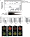
In wild-type mouse spermatocytes Spo11 transcripts peak past the stage in prophase in which meiotic DSBs are introduced. (A) Total Spo11 transcripts quantified in juvenile mice peak in late prophase I. Total Spo11 transcripts were quantified in testes of juvenile mice at 1 to 18 dpp. Total RNA was reverse transcribed and subjected to quantitative PCR (qPCR) using a TaqMan assay targeting the exon 11-12 boundary. Data were normalized to the 18-dpp sample. The scheme below the x axis indicates the predominant cell type corresponding to each age in wild-type juvenile mice, according to reference . Most prophase I mutants are arrested at stage IV (corresponding to early pachytene spermatocytes); therefore, their cell type composition would be comparable to that of a 13-dpp wild-type pup, whereas mutants arrested in metaphase I contain spermatocytes in all stages of prophase I. MR, meiotic replication; L, leptonema; Z, zygonema; EP, early pachynema; LP, late pachynema; D, diplonema. (B) Mutants arrested in prophase I display a 10-fold reduction in total Spo11 transcripts. Total Spo11 transcripts were quantified by reverse transcription-qPCR (RT-qPCR) in testes of several mutants arrested in prophase I. The ΔSpo11 strain was used as a negative control. Transcript levels for each homozygous mutant were normalized to those of its wild-type (wt) littermate. (C) Mutants arrested in metaphase I do not display the drastic reduction in total Spo11 transcript levels observed for prophase I mutants. Total Spo11 transcript levels in the testes of Mlh1/ mutant mice were compared to those of a wild-type littermate. (D) The low levels of total Spo11 transcripts detected in mutants arrested in prophase I are not a reflection of lower overall transcription levels due to the arrest. Cot-1 DNA consists of repetitive elements ubiquitously represented throughout the genome that can be used to detect nascent RNA transcripts in the nucleus by RNA FISH. The intensity of the nuclear Cot-1 signal correlates to the rate of transcription. Combined Cot-1 RNA FISH and SCP3 immunostaining were performed on wild-type and mutant spermatocyte spreads to assess overall transcription levels in leptotene and more advanced spermatocytes. Spermatocytes were classified as early (a, c, e, and g) or pachytene-like (b, d, f, and h) according to the SCP3 staining pattern.
FIG. 3.
FIG. 3.
Alternative splicing determines a switch from Spo11β to Spo11α transcripts in mid-prophase I. (A) Differential isoform-specific kinetics of transcription in juvenile mice. TaqMan assays targeting isoform-specific exon junctions were used to quantify Spo11α and -β transcripts by RT-qPCR in the testes of juvenile mice. Results were normalized to those for the 12-dpp sample. The table shows the relative ratios of mRNA levels (α/β). (B) Quantification of isoform-specific transcripts in isolated spermatocytes corroborates that Spo11α transcripts peak in late spermatocytes. Isoform-specific transcript levels were assessed by RT-qPCR on enriched cell populations isolated by FACS. Pie graphs show the percentages of spermatocytes undergoing the different stages of prophase in each cell population. Bar graphs show the relative levels of Spo11α and -β transcripts in each cell population normalized to those in P1. The table shows the relative ratios of mRNA levels (α/β). (C) Mutants arrested in prophase I contain only Spo11β transcripts, indicating that early spermatocytes synthesize solely Spo11β transcripts. Spo11α and -β transcripts were quantified by RT-qPCR in testes of different mutants arrested in prophase I or metaphase I. Bars represent the ratios of Spo11α or -β transcripts in homozygous mutants versus a wild-type littermate (KO/Wt).
FIG. 4.
FIG. 4.
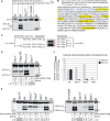
SPO11β polypeptide is expressed in early prophase I whereas SPO11α is only expressed once spermatocytes advance past early pachynema. (A) Generation of a monoclonal antibody (Ab) that can immunoprecipitate (Ip)/immunodetect SPO11 in mouse testis extracts. Total testis extracts (6 to 10 mg of total protein) from wild-type (wt) and Spo11/ mice were immunoprecipitated with anti-SPO11 (or nonspecific rabbit [R] IgG) and subsequently analyzed by Western blotting with the same antibody, anti-SPO11. M, molecular weight marker. (B) Presence of SPO11 in anti-SPO11 immunoprecipitates confirmed by mass spectrometry. Five peptides mapping to different areas of the protein (including exon 2 [blue]) identified SPO11 with 100% certainty in a wild-type extract immunoprecipitated with anti-SPO11 antibody (see Materials and Methods). The relatively low coverage (13%) did not allow identification of specific isoforms, given that a coverage above 95% would be required to identify SPO11α through the absence of peptides mapping to exon 2. (C) The estimated molecular masses of the proteins detected by the SPO11 antibody are in the range expected for SPO11 isoforms. Estimated molecular masses of the four bands detected by the antibody and expected molecular masses for the SPO11 isoforms annotated in Ensembl. (D) Testes from wild-type juvenile mice containing spermatocytes in early stages of prophase I contain primarily the SPO11β polypeptide. Testis extracts from juvenile mice at 11 dpp (early prophase I) and Dmc1/ and wild-type adult mice were immunoprecipitated/blotted with anti-SPO11 antibody. (E) Mutants arrested in midprophase express primarily the SPO11β polypeptide, whereas a mutant arrested in metaphase I expresses both isoforms. Total testis extracts from wild-type, Spo11/, Dmc1/, Hop2/, Atm/, and Mlh1/ mice (5 to 10 mg of total protein) were precipitated/blotted with anti-SPO11 antibody. Mutant testes are 0.2 to 0.5 the size of wild-type testes, so 3 or 4 mutant mice were used in order to immunoprecipitate extracts containing comparable amounts of total protein. The table specifies the number of mice used for each sample, the milligrams of total protein used for the Ip, and the α/β ratios. (F) Amount of SPO11 isoform precipitated from each extract normalized to total protein and to the wild-type extract. Shown is a comparison of the amounts of SPO11 isoform precipitated from mutant and wild-type extracts, taking into consideration the difference in total protein between extracts. For each isoform, the band intensity was divided by the amount of total protein in the extract and this ratio was divided by the corresponding ratio obtained for the wt sample. AU, arbitrary units.
FIG. 5.
FIG. 5.
SPO11β is expressed in excess such that a reduction by 50% in its level of expression does not translate into a comparable decline in the number of meiotic DSBs. (A) SPO11α and -β polypeptides are reduced by one-half in Spo11+/ testes, irrespective of ATM deficiency. Total testis extracts from Spo11+/+ and Spo11+/ littermates as well as Atm/ Spo11+/, Atm/ Spo11+/+, and Mlh1/ mice were immunoprecipitated/blotted with anti-SPO11 antibody. * and ** (also for panel B), minor splicing isoforms of SPO11α. (B) Amounts of SPO11 isoform precipitated from mutant and wild-type extracts, taking into consideration the difference in total protein between extracts. For each isoform, the band intensity (in arbitrary units) was divided by the amount of total protein in the extract. The normalized values for the α and β isoforms are documented in the table. (C) Amount of SPO11 isoform precipitated from each extract normalized to the wild-type (wt) extract. For each isoform, the band intensity (in arbitrary units) was divided by the amount of total protein in the extract and this ratio was divided by the corresponding ratio obtained for the wt sample: (intensity/mg total protein)mutant/(intensity/mg total protein)wt. (D) A 50% reduction in SPO11β polypeptide does not translate into a comparable decline in the number of DSBs. Spermatocyte spreads from Spo11+/+ and Spo11+/ (12-dpp) pups were stained for SCP3 and RAD51. The graph reports RAD51 focus numbers from 22 and 19 nuclei, respectively.
FIG. 6.
FIG. 6.
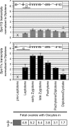
Oocytes synthesize Spo11α and -β transcripts during prophase I. TaqMan assays targeting isoform-specific exon junctions were used to quantify Spo11α and -β transcripts by RT-qPCR in fetal ovaries (E14.5 to E20/newborn females) carrying oocytes in the successive stages of prophase I. Results were normalized to those for the diplotene/dictyate oocyte sample. The table shows the relative ratios of mRNA levels (α/β).

Similar articles

Cited by

References

    1. Baker, S. M., A. W. Plug, T. A. Prolla, C. E. Bronner, A. C. Harris, X. Yao, D. M. Christie, C. Monell, N. Arnheim, A. Bradley, T. Ashley, and R. M. Liskay. 1996. Involvement of mouse Mlh1 in DNA mismatch repair and meiotic crossing over. Nat. Genet. 13:336-342. - PubMed
    1. Barchi, M., S. Mahadevaiah, M. Di Giacomo, F. Baudat, D. G. de Rooij, P. S. Burgoyne, M. Jasin, and S. Keeney. 2005. Surveillance of different recombination defects in mouse spermatocytes yields distinct responses despite elimination at an identical developmental stage. Mol. Cell. Biol. 25:7203-7215. - PMC - PubMed
    1. Barchi, M., I. Roig, M. Di Giacomo, D. G. de Rooij, S. Keeney, and M. Jasin. 2008. ATM promotes the obligate XY crossover and both crossover control and chromosome axis integrity on autosomes. PLoS Genet. 4:e1000076. - PMC - PubMed
    1. Barlow, C., S. Hirotsune, R. Paylor, M. Liyanage, M. Eckhaus, F. Collins, Y. Shiloh, J. N. Crawley, T. Ried, D. Tagle, and A. Wynshaw-Boris. 1996. Atm-deficient mice: a paradigm of ataxia telangiectasia. Cell 86:159-171. - PubMed
    1. Barlow, C., M. Liyanage, P. B. Moens, M. Tarsounas, K. Nagashima, K. Brown, S. Rottinghaus, S. P. Jackson, D. Tagle, T. Ried, and A. Wynshaw-Boris. 1998. Atm deficiency results in severe meiotic disruption as early as leptonema of prophase I. Development 125:4007-4017. - PubMed

Publication types

MeSH terms

LinkOut - more resources