Comparative Study
. 2010 Nov 11;116(19):3809-17.
doi: 10.1182/blood-2010-05-284141. Epub 2010 Jul 20.

Requirement of the human T-cell leukemia virus p12 and p30 products for infectivity of human dendritic cells and macaques but not rabbits

Affiliations
Comparative Study

Requirement of the human T-cell leukemia virus p12 and p30 products for infectivity of human dendritic cells and macaques but not rabbits

Valerio W Valeri et al. Blood. .

Abstract

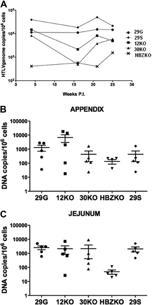
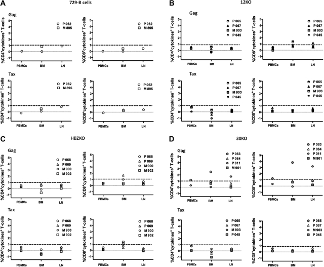
The identification of the genes necessary for human T-cell leukemia virus (HTLV-1) persistence in humans may provide targets for therapeutic approaches. We demonstrate that ablation of the HTLV-1 genes encoding p12, p30, or the HBZ protein, does not affect viral infectivity in rabbits and in this species, only the absence of HBZ is associated with a consistent reduction in virus levels. We observed reversion of the HTLV-1 mutants to the HTLV-1 wild-type genotype in none of the inoculated rabbits. In contrast, in macaques, the absence of HBZ was associated with reversion of the mutant virus to the wild-type genotype in 3 of the 4 animals within weeks from infection. Similarly, reversion to the wild type was observed in 2 of the 4 macaque inoculated with the p30 mutant. The 4 macaques exposed to the p12 knock remained seronegative, and only 2 animals were positive at a single time point for viral DNA in tissues. Interestingly, we found that the p12 and the p30 mutants were also severely impaired in their ability to replicate in human dendritic cells. These data suggest that infection of dendritic cells may be required for the establishment and maintenance of HTLV-1 infection in primate species.


Figures

Figure 1
Figure 1
Strategy for the ablation of p12, p30, and HBZ and characterization of the mutant viruses and cell lines. (A) Genetic organization of the HTLV-1 provirus genome and schematic representation of the overlapping orf I-IV (top). Amino acid changes in the mutant molecular clones (bottom). LTR indicates long terminal repeat. (B) Level of p19 Gag produced in the supernatants of the 729 B cell–infected cell lines measured by enzyme-linked immunoabsorbent assay. (C) Western blot analysis of the cell lysates from the 729 B-cell lines infected with the HTLV-1 mutant viruses with the use of antibodies to p24 Gag and to Tax. An antibody to tubulin was used as a control for equal loading of proteins. (D) Southern blot analysis of genomic DNA from the infected 729 B-cell lines. The numbers on the right represent the migration of the molecular weight (MW) marker.
Figure 2
Figure 2
12KO, 30KO, and HBZKO infectivity in primary human DCs. (A) The level of entry of HTLV ACH (WT), 30KO, 12KO, and HBZKO virions was determined by intracellular staining for the p19 Gag protein (white histogram) by fluorescence-activated cell sorting at 16 hours after virus exposure. The black histogram represents the staining of cells with the isotype control antibody. (B) Level of infection of DCs at 14 days after virus exposure, determined by the concentration of viral particles in the culture supernatant by the p19 Gag enzyme-linked immunoabsorbent assay. The results are presented as the percentage of HTLV-1 WT expression of the viral mutants. (C) Infection of DCs by 30KO in the presence (right) and absence (middle) of coexpression of the p30 c-DNA relative to WT HTLV (left), determined by the level of intracellular Tax at 1 week after infection. The black histogram refers to the antibody isotype control and the white histogram to the staining with the anti-Tax antibody.
Figure 3
Figure 3
Detection of HTLV-1 antigens by the sera of the infected rabbits. Sera from each rabbit infected with the HTLV-1 molecular clones were collected at weeks 0, 5, and 15 and reacted with Western blot strips carrying HTLV-1 antigens. All sera were negative in Western blot at week 0 (data not shown) but reacted with the p24 and p19 Gag proteins by weeks 5 and 15 from infection. The rgp46 refers to the recombinant peptide from the HTLV envelope protein; ns stands for a nonspecific HTLV-1 protein.
Figure 4
Figure 4
Virus level in the blood and tissues of the infected rabbits. Copies of HTLV-1 viral DNA per million of mononuclear cells in the blood of all infected animals overtime expresses as average (A) or presented for each animal at the time of killing in the appendix (B) and jejunum (C). The horizontal bars represent the average values. The statistical analysis of the data in blood was performed with the repeated measures of variance of the log-transformed copy numbers, and the P values for pairwise differences at each week were corrected by the Hochberg method.
Figure 5
Figure 5
Detection of HTLV-1 antigens by the sera of the infected macaques. Sera from each macaque exposed to the HTLV-1 molecular clones were collected at weeks 0, 2, 4, 8, and 11 and reacted with Western blot strips carrying HTLV-1 antigens. Macaque P062 and M985, which were exposed only to uninfected 729 B cells as control, did not seroconvert (A). None of the 12KO-inoculated macaques seroconverted to HTLV-1 antigens (B). All animals inoculated with the HBZKO either fully seroconverted (animals P068 and P069) or partially seroconverted (PM902 and M900) to HTLV-1 antigens (C); full seroconversion was observed only in animal P0011 inoculated with the 30KO virus (D).
Figure 6
Figure 6
HTLV-1 Tax and Gag–specific T-cell responses in macaques. Percentage of CD4+ T-cells (left) or CD8+T-cells (right) producing IL-2, tumor necrosis factor-α, or interferon-γ after stimulation with Gag (top) or Tax (bottom) overlapping peptides in control macaques (A), macaques exposed to 12KO (B) or HBZKO (C), and 30KO (D). All the data presented have been subtracted from the unstimulated cells background. The bolded dotted lines represent the background threshold of cytokine production observed in the control macaques. BM indicates bone marrow; LN, lymph node.

Similar articles

Cited by

References

    1. Poiesz BJ, Ruscetti FW, Gazdar AF, Bunn PA, Minna JD, Gallo RC. Detection and isolation of type C retrovirus particles from fresh and cultured lymphocytes of a patient with cutaneous T-cell lymphoma. Proc Natl Acad Sci U S A. 1980;77(12):7415–7419. - PMC - PubMed
    1. Gessain A, Barin F, Vernant J-C, et al. Antibodies to human T-lymphotropic virus type I in patients with tropical spastic paraparesis. Lancet. 1985;2(8452):407–410. - PubMed
    1. Ciminale V, Pavlakis GN, Derse D, Cunningham CP, Felber BK. Complex splicing in the human T-cell leukemia virus (HTLV) family of retroviruses: novel mRNAs and proteins produced by HTLV-I. J Virol. 1992;66(3):1737–1745. - PMC - PubMed
    1. Koralnik I, Gessain A, Klotman ME, Lo Monico A, Berneman ZN, Franchini G. Protein isoforms encoded by the pX region of the human T-cell leukemia/lymphotropic virus type I. Proc Natl Acad Sci U S A. 1992;89(18):8813–8817. - PMC - PubMed
    1. Lairmore MD, Franchini G. Human T-cell leukemia/lymphoma virus types 1 and 2. In: Knipe DM, Howley PM, editors. Fields Virology. 5th ed. Philadelphia, PA: Lippincott Williams & Wilkins; 2007. pp. 2071–2106.

Publication types

MeSH terms

Substances