Review
. 2010 Aug;11(8):539-51.
doi: 10.1038/nrn2870.

GSK3 signalling in neural development

Affiliations
Review

GSK3 signalling in neural development

Eun-Mi Hur et al. Nat Rev Neurosci. 2010 Aug.

Abstract

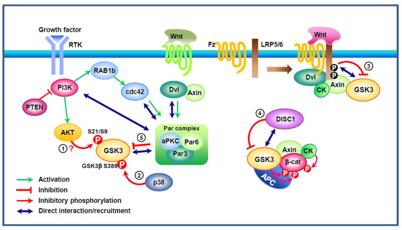
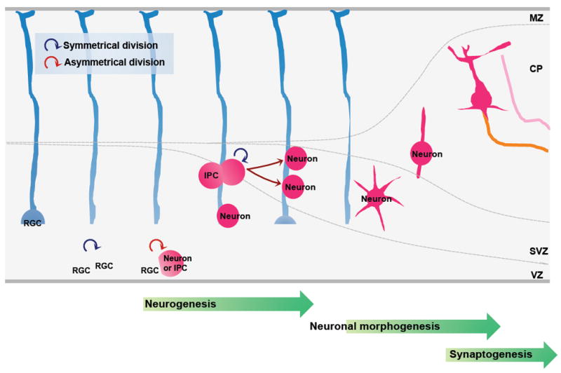
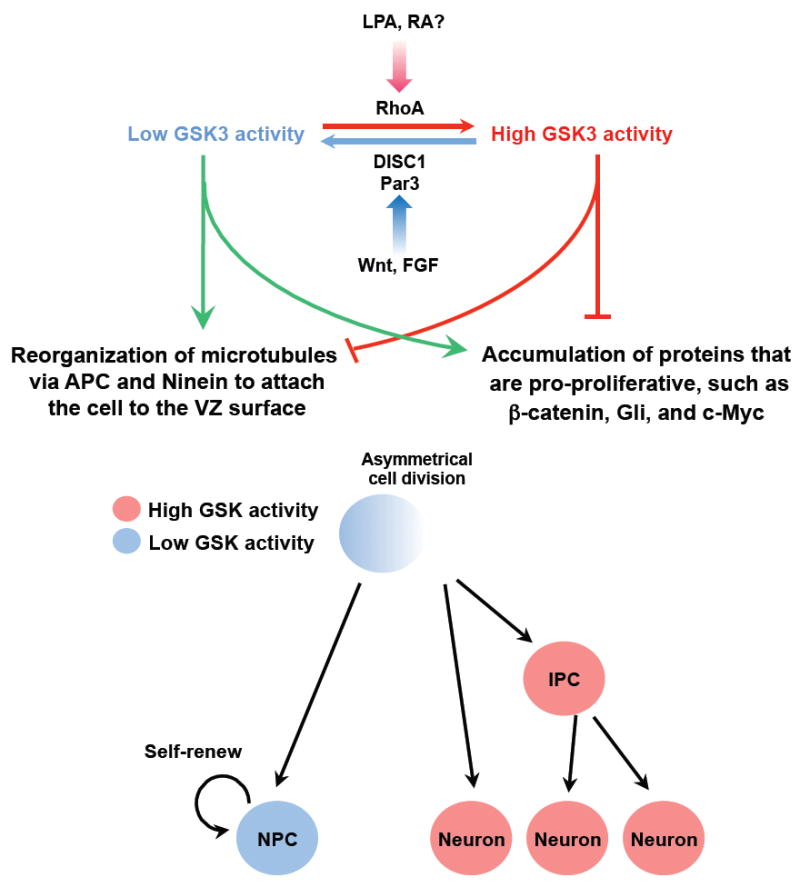
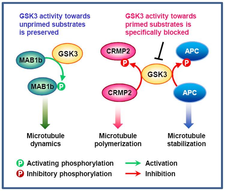
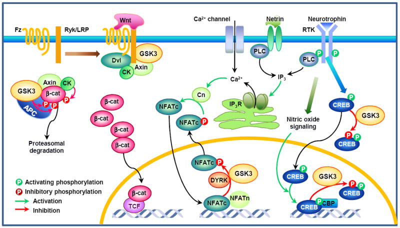
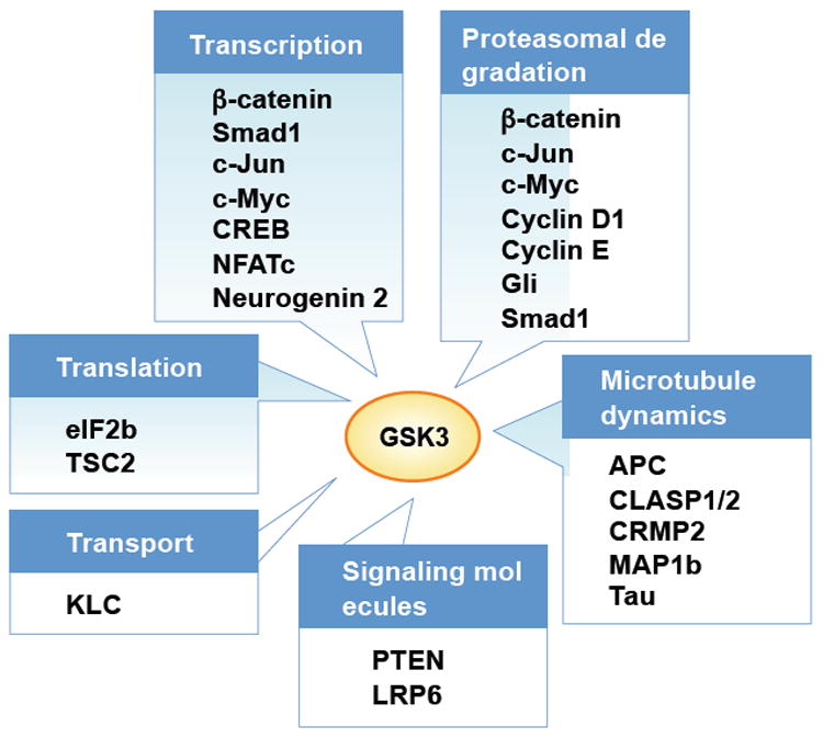
Recent evidence suggests that glycogen synthase kinase 3 (GSK3) proteins and their upstream and downstream regulators have key roles in many fundamental processes during neurodevelopment. Disruption of GSK3 signalling adversely affects brain development and is associated with several neurodevelopmental disorders. Here, we discuss the mechanisms by which GSK3 activity is regulated in the nervous system and provide an overview of the recent advances in the understanding of how GSK3 signalling controls neurogenesis, neuronal polarization and axon growth during brain development. These recent advances suggest that GSK3 is a crucial node that mediates various cellular processes that are controlled by multiple signalling molecules--for example, disrupted in schizophrenia 1 (DISC1), partitioning defective homologue 3 (PAR3), PAR6 and Wnt proteins--that regulate neurodevelopment.


Figures

Figure 1
Figure 1. Proposed models of glycogen synthase kinase (GSK) 3 inactivation
Upon activation of the phosphatidylinositol 3-kinase (PI3K) pathway downstream of receptor tyrosine kinase (RTK) signaling, AKT is thought to be the major kinase that inactivates GSK3 via phosphorylation of the N-terminal serine residue (S21 in GSK3α and S9 in GSK3β) (1). Phosphorylation of GSK3β (but not GSK3α) at S389 by p38 mitogen-activated protein kinase (MAPK) might be an alternative mechanism for inactivation (2). GSK3 regulation in the Wnt pathway is distinct from the way by which RTK signaling inhibits GSK3. In the canonical Wnt pathway, Wnt signaling recruits the destruction complex to the membrane, leading to the phosphorylation of the cytoplasmic tail of low-density lipoprotein receptor-related protein (LRP) 5/6 by casein kinase (CK) and GSK3β. Phosphorylated PPPSPXS motifs in the LRP5/6 intracellular domain directly inhibit GSK3’s activity towards β-catenin (3) , . In neuronal progenitor cells, a recent study showed that Disrupted in schizophrenia (DISC) 1 directly interacts with GSK3 and prevents it from phosphorylating β-catenin (4). Both in the Wnt and the RTK signaling pathways, GSK3 activity is also regulated by polarity proteins (5). GSK3 interacts with the Par complex — composed of Par3, Par6, and atypical protein kinase C (aPKC) — and so becomes phosphorylated and inactivated. Although phosphorylation of GSK3 occurs as a consequence, GSK3 phosphorylation is not required for its inactivation. β-cat, β-catenin; Dvl, dishevelled; Fz, Frizzled; PTEN, phosphatase and tensin homologue deleted on chromosome 10; RAP1b, Ras-related protein 1b.
Figure 2
Figure 2. Neural development of the mammalian neocortex
The formation of neural circuits during development involves the coordination of multiple cellular events, which can be divided into three steps. The first step is neurogenesis, during which a cohort of neural progenitors undergoes cycles of proliferation and differentiation to generate neurons in a highly regulated manner. The second step is neuronal morphogenesis, during which differentiated neurons migrate towards their final destination. While migrating, these neurons polarize to form axons and dendrites, which then grow and reach their specific target fields under the control of a myriad of guidance cues. The third step is synaptogenesis, during which axons cease growing and form synapses with their innervating targets. Radial glial cells (RGS) are progenitors that generate the majority of neurons in the developing neocortex, either directly or indirectly, by undergoing two phases of coordinated cell division. In the first, proliferative phase, which occurs early in development, they mainly divide symmetrically in the ventricular zone (VZ) to generate two similar progenitor cells, which can further self-renew to expand the progenitor pool. In the second, neurogenic phase, most of the radial glial cells divide asymmetrically to generate one self-renewing progenitor and one post-mitotic neuron or one intermediate progenitor cells (IPCs). The IPCs do not self-renew but migrate to the subventricular zone (SVZ) and divide symmetrically to form neurons. At the end of neurogenesis, some radial glial cells undergo a terminal symmetrical division to generate two neurons. The transition of progenitors from symmetrical to asymmetrical division and the asymmetrical division per se are tightly regulated to produce the final total number of neurons. Perturbation in any of these processes will cause defects in neurogenesis and cortical development. MZ, marginal zone; CP, cortical plate.
Figure 3
Figure 3. Proposed model for the role of GSK3 signaling during neurogenesis
During asymmetrical division of radial glial cells, the daughter cells may have different GSK3 activities via asymmetrical inheritance of upstream regulators (e. g. Par3, Disrupted in schizophrenia (DISC1), or Rho) under the control of extracellular factors (e. g. Wnt, fibroblast growth factor (FGF), lysophosphatidic acid (LPA), retinoic acid (RA)). The daughter cell with lower GSK3 activity accumulates pro-proliferation factors, such as β-catenin, Gli, and c-Myc. Microtubule-associated proteins adenomatosis polyposis coli (APC) and Ninein organize the astral microtubules to attach the cell to the ventricular zone (VZ) surface, and the cell adopts a progenitor fate. Conversely, the daughter cell with higher GSK3 activity removes pro-proliferative proteins and other proteins involved in microtubule assembly such as Ninein, presumably via the ubiquitin-proteasome system (UPS) mediated protein degradation. In addition, APC is unable to bind to the microtubules, disrupting the function of astral microtubules. As a result, the cell detaches from the VZ surface and become a neuron or an intermediate progenitor cell (IPC).
Figure 4
Figure 4. GSK3 in the regulation of neuronal polarization
Local activation of phosphatidylinositol 3-kinase (PI3K) at the tip of the nascent axon is thought to function as a landmark for the induction of neuronal polarization. PI3K-mediated production of phosphatidylinositol-3,4,5-trisphosphate (PIP3) leads to the activation of AKT and Ras-related protein 1b (RAP1b). RAP1b activates Cdc42, which recruits and activates the Par3/Par6/atypical protein kinase C (aPKC) complex, leading to the inactivation of GSK3, . Microtubule affinity-regulating kinase (MARK) 2 has also been suggested to function downstream of the Par complex to control neuronal polarization. aPKC in the Par complex associates with Dvl and mediates the Wnt5a-induced differentiation of axons. On the other hand, activation of AKT induces inhibitory phosphorylation of tuberous sclerosis complex (TSC) 2, by which its GTPase activating protein (GAP) activity towards Rheb is reduced, subsequently leading to the activation of mammalian target of rapamycin (mTOR) signaling. Although phosphorylation of TSC2 by AKT inhibits TSC1/2 activity, phosphorylation of TSC2 by GSK3 has an opposite effect. Thus, inhibition of GSK3 by PI3K or Wnt signaling would reduce GSK3-dependent stimulatory phosphorylation of TSC2 and thereby increase Rheb-GTP level, which in turn leads to the activation of mTOR-mediated translation. Activation of mTOR signaling in the axon induces local translation of collapsin response mediator protein (CRMP) 2 and Tau, substrates of GSK3. Activation of LKB1 by protein kinase A (PKA) or ribosomal S6 kinase (RSK) leads to the activation of SAD kinases, which in turn phosphorylate MAPs such as Tau, . The MAPs that are phosphorylated in response to LKB1-SAD kinase signaling to control neuronal polarization remain to be determined. A study in Xenopus shows that LKB1/XEEK1 physically associates with GSK3 and regulates its activity. Together with GSK3, MARK2 and SAD control microtubule assembly and dynamics by regulating the phosphorylation status of several MAPs. APC, adenomatosis polyposis coli; Dvl, dishevelled; 4EBP, eukaryotic translation initiation factor 4E-binding protein; ERK, extracellular signal regulated kinase; Fz, frizzled; GPCR, G protein-coupled receptor; MAP1b, microtubule-associated protein 1b; MEK, mitogen-activated protein kinase kinase; PTEN, phosphatase and tensin homologue deleted on chromosome 10; RTK, receptor tyrosine kinase; S6K, protein S6 kinase
Figure 5
Figure 5. Differential regulation of GSK3 substrates during axon growth
During rapid axon extension, GSK3 activity in the growth cone seems to be precisely controlled, so that its activity towards primed substrates is specifically blocked while its activity towards unprimed substrates is preserved. Inhibition of GSK3 activity towards collapsin response mediator protein (CRMP) 2 and adenomatosis polyposis coli (APC) allows CRMP2 and APC to bind microtubules, thereby increasing microtubule polymerization and stability, . By contrast, GSK3’s activity towards microtubule-associated protein (MAP) 1b is preserved in the growth cone. Phosphorylation of MAP1b maintains microtubules in a dynamic state, which is essential for axon growth, . In this way, GSK3 can coordinate essential properties of axonal microtubules to ensure optimal microtubule assembly in axons.
Figure 6
Figure 6. Potential roles of GSK3 in the transcriptional regulation of axon growth
In the absence of Wnt, the transcriptional co-activator β-catenin (β-cat) is phosphorylated by GSK3 in the destruction complex, which tags β-cat for proteasomal degradation. Wnt promotes the association of Frizzled (Fz) receptor and the co-receptor low-density lipoprotein receptor-related protein (LRP) or Ryk, leading to recruitment of the cytoplasmic dishevelled (Dvl) and the destruction complex to the membrane. Inhibition of GSK3 activity results in the accumulation of hypophosphorylated β-cat, which then can enter the nucleus and promote T-cell factor (TCF, also known as lymphoid enhancer-binding factor)-mediated gene transcription. Another family of transcription factors involved in axon growth is nuclear factor of activated T-cells (NFAT) proteins. Neurotrophins and netrins require calcineurin (Cn)/NFAT signaling for axon growth. A rise in intracellular Ca2+ induced by neurotrophins, netrins, or calcium channels activates the serine/threonine phosphatase Cn, which dephosphorylates NFATc proteins. Dephosphorylation of NFATc triggers their nuclear translocation, where they form complexes with NFATn to induce gene transcription. NFATc proteins are rapidly excluded from the nucleus through sequential phosphorylation, first by a priming kinase such as protein kinase A (PKA) and dual-specificity tyrosine-phosphorylation regulated kinase (DYRK) 1A, then by GSK3. Because NFAT phosphorylation by GSK3 inhibits its DNA binding activity and is required for its nuclear export, GSK3 could play a role in NFAT-mediated gene transcription downstream of neurotrophin and/or netrin signaling. cAMP response element binding protein (CREB) is a well-established transcription factor that mediates neurotrophin-induced axon growth. Neurotrophins and other stimuli lead to the phosphorylation of CREB at S133, which allows CREB binding to the transcriptional co-activator CREB-binding protein (CBP). In developing neurons, DNA binding activity of CREB is tightly controlled via nitric oxide signaling, as opposed to the classical model in which CREB is constitutively bound to promoter sequences. Similar to NFATc, GSK phosphorylation of CREB prevents it from binding to DNA. Because GSK3 is found in the cytoplasm and the nucleus, CREB phosphorylation can occur in both places. However, it should be noted that GSK regulation of NFAT and CREB has not been directly tested in the context of axon growth. APC, adenomatous polyposis coli; CK, casein kinase; IP3, inositol-1,4,5-trisphosphate; IP3R, IP3 receptor.
Figure 7
Figure 7. Representative substrates of GSK3 implicated in neural development
GSK3 regulation of neural development can be achieved through phosphorylation of proteins that control key steps in neurodevelopment. GSK3 phosphorylates several transcription factors, such as β-catentin, Smad1, c-Jun, c-Myc, cAMP response element binding protein (CREB), nuclear factor of activated T-cells (NFATc), and neurogenin 2, many of which undergo proteasomal degradation after GSK3 phosphorylation. GSK3 phosphorylation mediates proteasomal targeting and degradation of other proteins as well, such as cyclin D1 and cyclin E. The most well-characterized substrates of GSK3 involved in neural development are microtubule-associated proteins that control neuronal polarization and axon growth. These substrates include adenomatosis polyposis coli (APC), CLIP-associated protein (CLASP) 1 and 2, collapsin response mediator protein (CRMP) 2, MAP (microtubule-associated protein) 1b and Tau. Although CLASP is enriched in the growth cone and mediate Slit-induced axon repulsion during neural development in Drosophila, its localization and role during neuronal polarization and/or axon growth in the mammalian nervous system remains to be determined. Signaling molecules such as phosphatase and tensin homologue deleted on chromosome 10 (PTEN) and Wnt co-receptor low-density lipoprotein receptor-related protein (LRP) 6 are also phosphorylated by GSK3. Other examples are kinesin light chain (KLC) that regulates selective transport, and essential components of the translational machinery, such as eukaryotic initiation factor (eIF) 2b and tuberous sclerosis complex (TSC2).

Similar articles

Cited by

References

    1. Woodgett JR, Cohen P. Multisite phosphorylation of glycogen synthase. Molecular basis for the substrate specificity of glycogen synthase kinase-3 and casein kinase-II (glycogen synthase kinase-5) Biochim Biophys Acta. 1984;788:339–47. - PubMed
    1. Wang Y, Roach PJ. Inactivation of rabbit muscle glycogen synthase by glycogen synthase kinase-3. Dominant role of the phosphorylation of Ser-640 (site-3a) J Biol Chem. 1993;268:23876–80. - PubMed
    1. Woodgett JR. Molecular cloning and expression of glycogen synthase kinase-3/factor A. EMBO J. 1990;9:2431–8. - PMC - PubMed
    1. Mukai F, Ishiguro K, Sano Y, Fujita SC. Alternative splicing isoform of tau protein kinase I/glycogen synthase kinase 3beta. J Neurochem. 2002;81:1073–83. - PubMed
    1. Castano Z, Gordon-Weeks PR, Kypta RM. The neuron-specific isoform of glycogen synthase kinase-3beta is required for axon growth. J Neurochem. 2010;113:117–30. - PubMed

Publication types

MeSH terms