Review
. 2010 Aug;11(8):563-76.
doi: 10.1038/nrn2880.

Transcriptional regulation of photoreceptor development and homeostasis in the mammalian retina

Affiliations
Review

Transcriptional regulation of photoreceptor development and homeostasis in the mammalian retina

Anand Swaroop et al. Nat Rev Neurosci. 2010 Aug.

Abstract

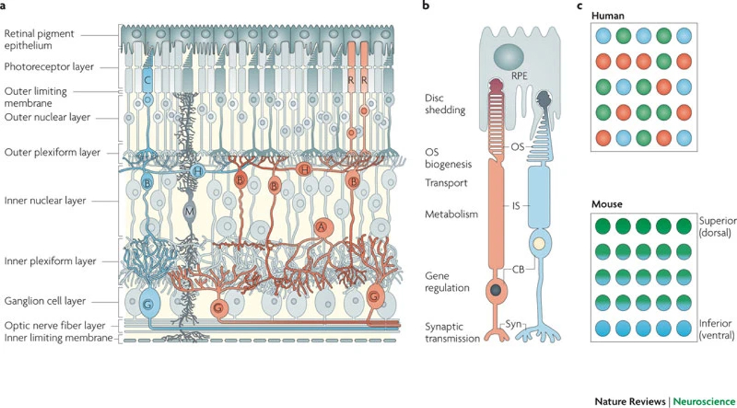
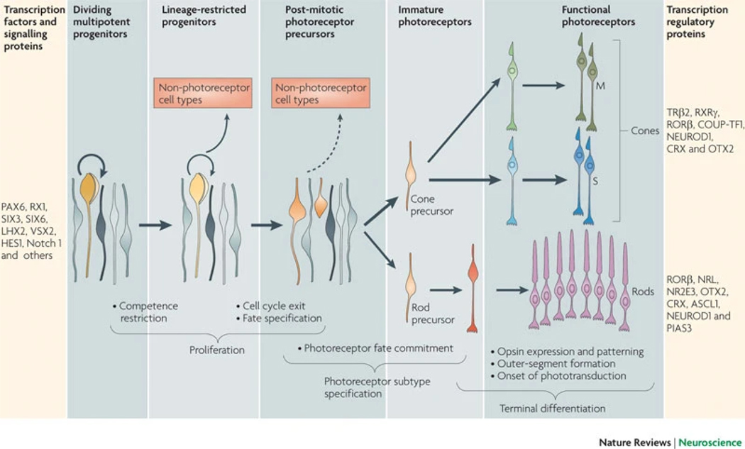
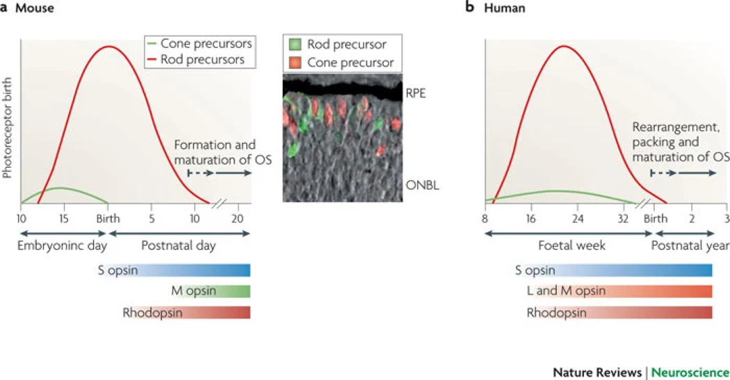
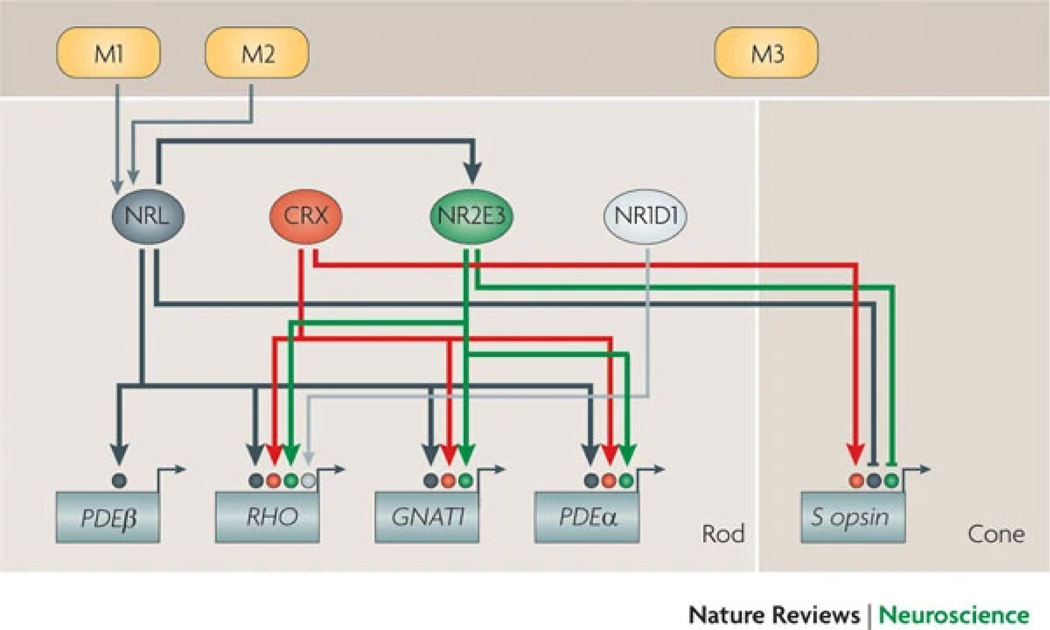
In the developing vertebrate retina, diverse neuronal subtypes originate from multipotent progenitors in a conserved order and are integrated into an intricate laminated architecture. Recent progress in mammalian photoreceptor development has identified a complex relationship between six key transcription-regulatory factors (RORbeta, OTX2, NRL, CRX, NR2E3 and TRbeta2) that determine rod versus M cone or S cone cell fate. We propose a step-wise 'transcriptional dominance' model of photoreceptor cell fate determination, with the S cone representing the default state of a generic photoreceptor precursor. Elucidation of gene-regulatory networks that dictate photoreceptor genesis and homeostasis will have wider implications for understanding the development of nervous system function and for the treatment of neurodegenerative diseases.


Conflict of interest statement

Competing interests statement

The authors declare no competing financial interests.

Figures

Figure 1:
Figure 1:. Functional circuitry of the retina.
a | Organization of retinal circuits. Rod (R) and cone (C) photoreceptors have cell bodies (CBs) in the outer nuclear layer and extend inner segments (IS), which contain metabolic machinery, and outer segments (OS), which associate with retinal pigment epithelial (RPE) cells. Photoreceptor axons terminate in the outer plexiform layer and synapse with horizontal (H) and bipolar (B) cells in the inner nuclear layer, which also contains Müller glial (M) and amacrine (A) cells. Bipolar cells relay signals to amacrine and ganglion (G) cells through synapses in the inner plexiform layer. Ganglion cell axons project towards the optic nerve head and carry signals to the brain. End-feet of Müller glia form the outer and inner limiting membranes. A representative cone pathway is shown in blue and a representative rod pathway is shown in orange. b | Illustrations of rod and cone morphologies that include subcellular locations of functions. c | A surface-view representation of cone distribution across the mammalian retina. In humans, cones express S opsins (peak sensitivity to blue light), L opsins (peak sensitivity to red light) or M opsins (peak sensitivity to green light) in a mosaic-like pattern. In mice, cones express S opsins and M opsins in opposing distribution gradients along the superior (M opsin-high) to inferior (S opsin-high) axis. Syn, synaptic terminus.
Figure 2:
Figure 2:. Stages of photoreceptor development.
Early in retinogenesis, multipotent retinal progenitor cells (RPCs) divide and produce additional multipotent progenitors (thick circular arrow) or progenitor cells that become restricted in their competence to generate various cell types (thin circular arrow). Some of these proliferating cells become restricted to a lineage that will give rise to at least one photoreceptor cell and possibly to non-photoreceptor cells. After cell cycle exit, postmitotic precursors can remain plastic. During cell type specification of photoreceptors, precursors are directed to become cones or rods that eventually express photopigments (M opsin and S opsin in cones, and rhodopsin in rods), and form outer segments and synapses. The bar on the far left lists key transcription factors and signalling proteins that maintain RPC multipotency and proliferation: paired box protein PAX6, retinal homeobox protein RX1, SIX3, SIX6, LIM–homeobox protein LHX2, visual system homeobox 2 (VSX2), HES1 and Notch 1. The bar on the far right lists key transcription regulatory proteins that are involved in cone and rod differentiation and maintenance (for cones: thyroid hormone receptor β2 (TRβ2), retinoid X receptor-γ (RXRγ), nuclear receptor RORβ, COUP transcription factor 1 (COUP-TF1; also known as NR2F1), neurogenic differentiation factor 1 (NEUROD1), cone–rod homeobox protein (CRX) and homeobox protein OTX2; for rods: RORβ, neural retina leucine zipper protein (NRL), photoreceptor-specific nuclear receptor (NR2E3), OTX2, CRX, achaete-scute homologue 1 (ASCL1; also known as MASH1), NEUROD1 and E3 SUMO-protein ligase PIAS3).
Figure 3:
Figure 3:. Photoreceptor genesis and maturation in mice and humans.
The relative numbers of cone and rod precursor cells that are born over time are shown for mouse and human retinogenesis. a | In mice, cones are generated prenatally as early as embryonic day (E) 11. Rods, which vastly outnumber cones, are generated both before and after birth, from around E12 to postnatal day (P) 10. Expression of S opsin begins at ~E18, that of M opsin at ~P6 and that of rhodopsin at ~P2. Photopigment levels increase substantially until after weaning in mice. As photoreceptors mature, outer segments (OS) and synapses form. The inset panel shows double fluorescent detection of cone (thyroid hormone receptor β2-positive cells (red staining)) and rod precursors (neural retina leucine zipper protein (NRL) -positive cells (green staining)) in mouse retina at E18, superimposed on a phase contrast picture that reveals the outer neuroblastic layer (ONBL) (D. Sharlin, Alok Swaroop and D. Forrest, unpublished data). Newly generated photoreceptors tend to reside near the edge of the retina. b | In humans, cones and rods are generated around foetal week (Fwk) 8 and Fwk 10, respectively. Generation of cones is completed prenatally, whereas that of rods continues into the early postnatal period. Expression of S opsin is observed at ~Fwk 12, and that of L opsin, M opsin and rhodopsin at ~ Fwk 15. Functional maturation of photoreceptors continues postnatally. RPE, retinal pigment epithelium. Data are taken from Refs , , , , , , .
Figure 4:
Figure 4:. Transcriptional dominance model of photoreceptor cell fate determination.
A generic photoreceptor is formed under the control of homeobox protein OTX2 and other undetermined signals. This precursor is programmed to possess a ‘default’ S cone state under the control of OTX2 (and/or cone–rod homeobox protein (CRX)) and nuclear receptor RORβ unless diverted into a rod or M cone state by additional signals. The ‘NRL control box’ determines whether a precursor becomes a rod or a cone, and the ‘thyroid hormone receptor β2 (TRβ2) control box’ prompts a cone to acquire an M opsin or an S opsin identity. Each control box is subject to modifying regulators. Induction of neural retina leucine zipper protein (NRL) and its target, photoreceptor-specific nuclear receptor (NR2E3), induces a rod state and suppresses cone genes, which consolidates the rod fate. Other factors involved in rod development include E3 SUMO-protein ligase PIAS3, neurogenic differentiation factor 1 (NEUROD1), achaete-scute homologue 1 (ASCL1; also known as MASH1), myocyte enhancer factor 2C and retinoblastoma-associated protein, probably at both early and later stages of differentiation. If NRL and NR2E3 fail to act, photoreceptor precursors follow the ‘default’ pathway to S cones unless TRβ2 and its ligand triiodothyronine (T3) induce M opsin and suppress S opsin expression in spatially restricted patterns in cone subpopulations over the retina. Other factors involved in M opsin and S opsin patterning include retinoid X receptor-γ (RXRγ), RORβ, RORα, COUP transcription factors (COUP-TFs) and CRX. NEUROD1 and bone morphogenetic protein receptor type 1A–1B contribute to inducing the expression of TRβ2 and COUP-TF, respectively.
Figure 5:
Figure 5:. A simplified representation of rod gene-regulatory networks.
Neural retina leucine zipper protein (NRL) acts synergistically with cone–rod homeobox protein (CRX) and/or photoreceptor-specific nuclear receptor (NR2E3) in many distinct complexes that bind to enhancer sequences and dictate the expression of various rod genes. The rhodopsin (RHO) promoter is likely to be regulated by additional factors, such as nuclear receptor subfamily 1 group D member 1 (NR1D1), to maintain high but precisely controlled levels in rods. Other promoters (such as that for rod cGMP-specific 3′,5′-cyclic phosphodiesterase subunit-β (PDEβ)) may bind the widely expressed transcription factors SP1 and SP4 (Ref. 153), together with NRL. NRL and NR2E3 repress the activity of cone genes (such as S opsin), whereas CRX activates both rod and cone genes. M1, M2 and M3 indicate modulators (such as fibroblast growth factor 2, retinoic acid and post-translational modifiers) that control or fine-tune the activity of key transcription factors. Thin lines denote experiment-based suggested links, whereas thick lines denote confirmed regulatory connections. GNAT1, guanine nucleotide-binding protein G(t) subunit α1.
None

References

    1. Rodieck RW The First Steps in Seeing (Sinauer Associates Publishers, Sunderland, Massachusetts,1998).
    1. Dowling JE The Retina: An Approachable Part of the Brain. (Belknap Press, Harvard Univ. Press, 1987).
    1. Masland RH The fundamental plan of the retina. Nature Neurosci. 4, 877–886 (2001). - PubMed
    1. Wassle H. Parallel processing in the mammalian retina. Nature Rev. Neurosci. 5, 747–757 (2004). - PubMed
    1. Luo DG, Xue T. & Yau KW How vision begins: an odyssey. Proc. Natl Acad. Sci. USA 105, 9855–9862 (2008). - PMC - PubMed

Publication types

LinkOut - more resources