

Ochratoxin A: in utero exposure in mice induces adducts in testicular DNA
- PMID: 20648226
- PMCID: PMC2905807
- DOI: 10.3390/toxins2061428
Ochratoxin A: in utero exposure in mice induces adducts in testicular DNA
Abstract
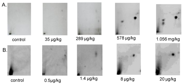
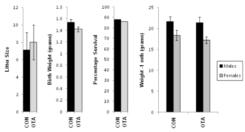
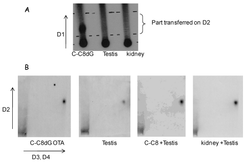
Ochratoxin A (OTA) is a nephrotoxin and carcinogen that is associated with Balkan endemic nephropathy and urinary tract tumors. OTA crosses the placenta and causes adducts in the liver and kidney DNA of newborns. Because the testis and kidney develop from the same embryonic tissue, we reasoned that OTA also may cause adducts transplacentally in the testis. We tested the hypothesis that acute exposure to OTA, via food and via exposure in utero, causes adducts in testicular DNA and that these lesions are identical to those that can be produced in the kidney and testis by the consumption of OTA. Adult mice received a single dose of OTA (from 0–1,056 µg/kg) by gavage. Pregnant mice received a single i.p. injection of OTA (2.5 mg/kg) at gestation day 17. DNA adducts were determined by 32P-postlabeling. Gavage-fed animals sacrificed after 48 hours accumulated OTA in kidney and testis and showed DNA adducts in kidney and testis. Some OTA metabolites isolated from the tissues were similar in both organs (kidney and testis). The litters of mice exposed prenatally to OTA showed no signs of overt toxicity. However, newborn and 1-month old males had DNA adducts in kidney and testis that were chromatographically similar to DNA adducts observed in the kidney and testis of gavage-fed adults. One adduct was identified previously as C8-dG-OTA adduct by LC MS/MS. No adducts were observed in males from dams not exposed to OTA. Our findings that in utero exposure to OTA causes adducts in the testicular DNA of male offspring support a possible role for OTA in testicular cancer.
Keywords: DNA adduct; epidemiology; ochratoxin; testicular cancer; transplacental contamination.
Figures


Comment in
-
Comments on "Ochratoxin A: In utero Exposure in Mice Induces Adducts in Testicular DNA. Toxins 2010, 2, 1428-1444"-Mis-Citation of Rat Literature to Justify a Hypothetical Role for Ochratoxin A in Testicular Cancer.Toxins (Basel). 2010 Oct;2(10):2333-6; author reply 2337-9. doi: 10.3390/toxins2102333. Epub 2010 Sep 29. Toxins (Basel). 2010. PMID: 22069555 Free PMC article.
References
-
- O’Brien E., Dietrich D.R. Ochratoxin A: The continuing enigma. Critical Reviews. Toxicology. 2005;35:33–60. - PubMed
-
- Pfohl-Leszkowicz A., Manderville R.A. Ochratoxin A: An overview on toxicity and carcinogenicity in animals and humans. Mol. Nutr. Food Res. 2007;51:61–99. - PubMed
-
- Krogh P. Epidemiology of mycotoxic porcine nephropathy. Nord. Vet. Med. 1976;28:452–458. - PubMed
-
- Stoev S.D. The role of ochratoxin A as a possible cause of Balkan endemic nephropathy and its risk evaluation. Vet. Human Toxicol. 1998;40:352–360. - PubMed
Publication types
MeSH terms
Substances
Grants and funding
LinkOut - more resources
Full Text Sources
