. 2010 Feb;1(1):2-26.
doi: 10.1159/000276766. Epub 2010 Jan 15.

Noonan syndrome: clinical aspects and molecular pathogenesis

Affiliations

Noonan syndrome: clinical aspects and molecular pathogenesis

M Tartaglia et al. Mol Syndromol. 2010 Feb.

Abstract

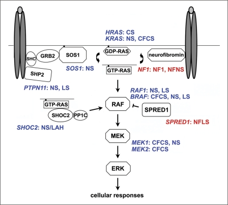
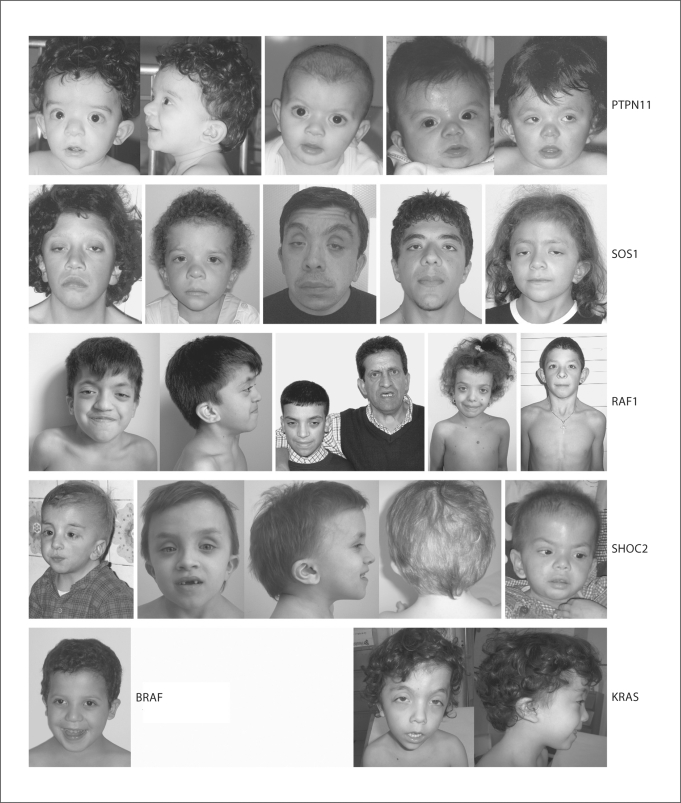
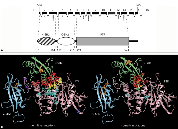
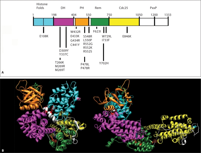
Noonan syndrome (NS) is a relatively common, clinically variable and genetically heterogeneous developmental disorder characterized by postnatally reduced growth, distinctive facial dysmorphism, cardiac defects and variable cognitive deficits. Other associated features include ectodermal and skeletal defects, cryptorchidism, lymphatic dysplasias, bleeding tendency, and, rarely, predisposition to hematologic malignancies during childhood. NS is caused by mutations in the PTPN11, SOS1, KRAS, RAF1, BRAF and MEK1 (MAP2K1) genes, accounting for approximately 70% of affected individuals. SHP2 (encoded by PTPN11), SOS1, BRAF, RAF1 and MEK1 positively contribute to RAS-MAPK signaling, and possess complex autoinhibitory mechanisms that are impaired by mutations. Similarly, reduced GTPase activity or increased guanine nucleotide release underlie the aberrant signal flow through the MAPK cascade promoted by most KRAS mutations. More recently, a single missense mutation in SHOC2, which encodes a cytoplasmic scaffold positively controlling RAF1 activation, has been discovered to cause a closely related phenotype previously termed Noonan-like syndrome with loose anagen hair. This mutation promotes aberrantly acquired N-myristoylation of the protein, resulting in its constitutive targeting to the plasma membrane and dysregulated function. PTPN11, BRAF and RAF1 mutations also account for approximately 95% of LEOPARD syndrome, a condition which resembles NS phenotypically but is characterized by multiple lentigines dispersed throughout the body, café-au-lait spots, and a higher prevalence of electrocardiographic conduction abnormalities, obstructive cardiomyopathy and sensorineural hearing deficits. These recent discoveries demonstrate that the substantial phenotypic variation characterizing NS and related conditions can be ascribed, in part, to the gene mutated and even the specific molecular lesion involved.

Keywords: Cardiofaciocutaneous syndrome; Costello syndrome; Genotype-phenotype correlations; LEOPARD syndrome; Molecular basis of disease; Molecular epidemiology; Mutation analysis; Neurocardiofacialcutaneous syndrome family; Noonan syndrome; RAS signaling.


Figures

Fig. 1
Fig. 1
Schematic diagram showing the RAS-MAPK signal transduction pathway and affected disease genes in disorders of the neurocardiofacialcutaneous syndrome family. The double ovals in dark grey and the light grey ovals represent generic dimerized cell-surface receptors binding to their ligand. Abbreviations: CFCS, cardiofaciocutaneous syndrome; CS: Costello syndrome; LS, LEOPARD syndrome; NF1, neurofibromatosis type 1; NFLS, neurofibromatosis type 1-like syndrome (also termed Legius syndrome); NFNS, neurofibromatosis-Noonan syndrome; NS, Noonan syndrome; NS/LAH, Noonan-like syndrome with loose anagen hair.
Fig. 2
Fig. 2
Dysmorphic facial features in Noonan syndrome. Series of affected individuals heterozygous for mutations in different disease genes are shown.
Fig. 3
Fig. 3
PTPN11 gene organization, SHP2 domain structure and location of affected residues in human disease. A The PTPN11 gene and its encoded protein. The numbered, filled boxes at the top indicate the coding exons; the positions of the ATG and TGA codons are shown. Exon location of disease-associated mutations is indicated (the number of asterisks is an index of the relative prevalence of mutations within each exon). SHP2's functional domains, consisting of 2 tandemly arranged SH2 domains at the N-terminus (N-SH2 and C-SH2) followed by a protein tyrosine phosphatase (PTP) domain, are shown below. The numbers below that cartoon indicate the amino acid boundaries of those domains. B Location of mutated residues in the 3-dimensional structure of SHP2 in its catalytically inactive conformation. Residues affected by germline (left) or somatically acquired (right) mutations are shown with their lateral chains colored according to the classification proposed by Tartaglia et al. [2006] (red, mutations affecting the N-SH2/PTP interaction; yellow, mutations affecting the N-SH2/PTP interaction and possibly catalysis; green, mutations affecting the N-SH2/PTP interaction and possibly substrate specificity; cyan, mutations affecting the N-SH2/PTP interaction and/or catalysis; orange, mutations promoting increased SH2 phosphopeptide-binding affinity or affecting specificity; violet, mutations affecting SH2 orientation or mobility; blue, unclassified).
Fig. 4
Fig. 4
SOS1 domain structure and location of affected residues in NS. A SOS1 missense mutations are positioned below the cartoon of the SOS1 protein with its functional domains indicated above. Abbreviations: DH, Dbl homology domain; PH, plekstrin homology domain; Rem, RAS exchanger motif; PxxP, proline-rich motif. B Location of the mutated residues on the 3-dimensional structure of SOS1. The functional domains are color-coded as follows: cyan, histone folds; magenta, DH; orange, PH; green, Rem; yellow, Cdc25. Residues affected by mutations are indicated with their lateral chains (violet, histone folds; blue, DH; green, PH; red, helical linker; orange, Rem; cyan, Cdc25). Based on Sondermann et al. [2005], who utilized structural data and computational modeling.
Fig. 5
Fig. 5
KRAS gene organization, protein domain structure and location of affected residues specifically affecting the KRASB isoform. A KRAS gene organization and transcript processing to produce the alternative KRAS isoforms A and B. The numbered black and grey boxes indicate the invariant coding exons and exons undergoing alternative splicing, respectively. KRASB mRNA results from exon 5 skipping. In KRASA mRNA, exon 6 encodes the 3′-UTR. The arrows indicate location of mutations affecting codons 152, 153 and 156. B Schematic diagram (above) and tridimensional representation (below) of the structural and functional domains defined within RAS proteins. The motifs required for signaling function (PM1 to PM3 indicate residues involved in binding to the phosphate groups, while G1 to G3 are those involved in binding to the guanine base) are indicated. The hypervariable region is shown in grey, together with the C-terminal motifs that direct post-translational processing and plasma membrane anchoring (dark grey). The GTP/GDP binding pocket is shown in cyan (guanine ring-binding surface) and yellow (triphosphate group binding surface) together with the Switch I (green) and Switch II (magenta) domains, according to the GTP-bound RAS conformation.
Fig. 6
Fig. 6
RAF1 and BRAF domain structures and location of affected residues in human disease. The domains of the RAF1 (A) and BRAF (B) proteins are indicated (CR, conserved region; RBD, RAS-binding domain; CRD, cysteine-rich domain). Germline (RAF1: NS and LS; BRAF: NS, LS and CFCS) and somatic (associated with cancer) mutations are reported above and below each cartoon, respectively. Only somatic BRAF mutations with prevalence ≥1.5%, according the COSMIC database (http://www.sanger.ac.uk/genetics/CGP/cosmic/) are reported. The BRAF T599I substitution, which rarely occurs in cancer and is homologous to the NS-causing RAF1 T491I change, is also reported. BRAF missense changes associated with a phenotype fitting NS or LS are colored blue and orange, respectively.
Fig. 7
Fig. 7
The disease-causing c.4A>G change in SHOC2 promotes protein myristoylation and cell membrane targeting. A SHOC2 genomic organization and protein structure. The coding exons are shown at the top as numbered filled boxes. Intronic regions are reported as dotted lines. SHOC2 motifs comprise an N-terminal lysine-rich region (grey colored) followed by 19 leucine-rich repeats. Numbers above the domain structure indicate the amino acid boundaries of those domains. B Confocal laser scanning microscopy analysis documents that SHOC2wt (red) is uniformly distributed in the cytoplasm and nucleus in starved cells (left), while SHOC2S2G (red) is specifically targeted to the cell membrane. Actin cytoskeleton (green) and nuclei (blue) are also shown.

References

    1. Adekunle O, Gopee A, el-Sayed M, Thilaganathan B. Increased first trimester nuchal translucency: Pregnancy and infant outcomes after routine screening for Down's syndrome in an unselected antenatal population. Br J Radiol. 1999;72:457–460. - PubMed
    1. Adjei AA. Blocking oncogenic Ras signaling for cancer therapy. J Natl Cancer Inst. 2001;93:1062–1074. - PubMed
    1. Ahmed ML, Foot AB, Edge JA, Lamkin VA, Savage MO, Dunger DB. Noonan's syndrome: Abnormalities of the growth hormone/IGF axis and the response to treatment with human biosynthetic growth hormone. Acta Paediatr Scand. 1991;80:446–450. - PubMed
    1. Alfieri P, Cesarini L, Zampino G, Pantaleoni F, Selicorni A, et al. Visual function in Noonan and LEOPARD syndrome. Neuropediatrics. 2008;39:335–340. - PubMed
    1. Allanson JE. Noonan syndrome. Am J Med Genet C Semin Med Genet. 2007;145C:274–279. - PubMed