. 2010 Dec;16(12):3719-26.
doi: 10.1089/ten.TEA.2009.0836. Epub 2010 Aug 30.

Adipose stroma induces branching morphogenesis of engineered epithelial tubules

Affiliations

Adipose stroma induces branching morphogenesis of engineered epithelial tubules

Amira L Pavlovich et al. Tissue Eng Part A. 2010 Dec.

Abstract

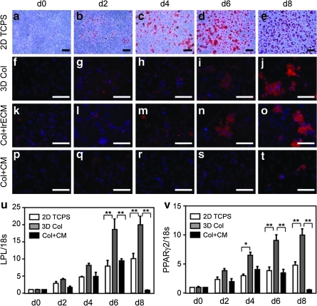
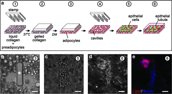
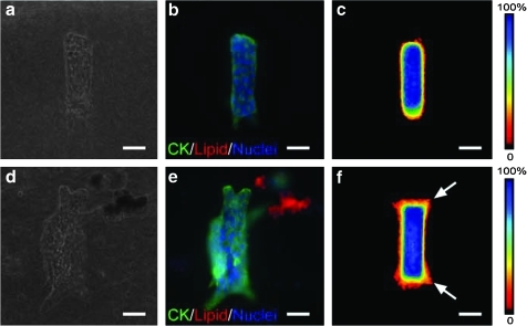
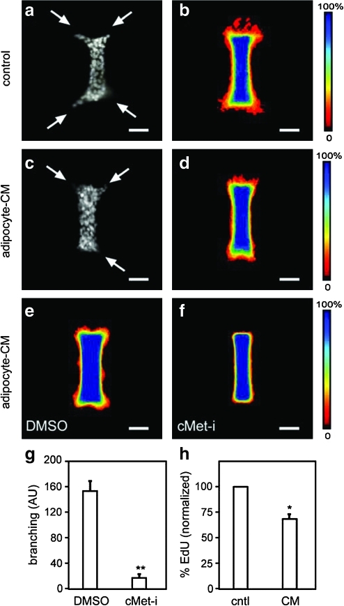
The mammary gland and other treelike organs develop their characteristic fractal geometries through branching morphogenesis, a process in which the epithelium bifurcates and invades into the surrounding stroma. Controlling the pattern of branching is critical for engineering these organs. In vivo, the branching process is instructed by stromal-epithelial interactions and adipocytes form the largest component of the fatty stroma that surrounds the mammary epithelium. Here, we used microlithographic approaches to engineer a three-dimensional culture model that enables analysis of the effect of adipocytes on the pattern of branching morphogenesis of mammary epithelial cells. We found that adipocyte-rich stroma induces branching through paracrine signals, including hepatocyte growth factor, but does not affect the branching pattern per se. This tissue engineering approach can be expanded to other organs, and should enable piecemeal analysis of the cellular populations that control patterning during normal development.


Figures

FIG. 1.
FIG. 1.
Time course of adipogenesis. (a–e) Oil red O staining of 3T3-L1 preadipocytes induced to differentiate on two-dimensional (2D) tissue culture polystyrene (TCPS). Sudan III (red) and nuclear (blue) staining of 3T3-L1 cells induced to differentiate in (f–j) three-dimensional collagen (3D Col), (k–o) collagen plus 5% laminin-rich extracellular matrix (Col + lrECM), or (p–t) collagen in the presence of the mammary epithelial-conditioned medium (CM; Col + CM). Extent of differentiation as determined by quantitative reverse transcription (RT)/polymerase chain reaction analysis of (u) lipoprotein lipase (LPL) and (v) peroxisome proliferator-activated receptor (PPAR)-γ2 expression levels; shown are average and standard error of the mean (SEM). *p < 0.01; **p < 0.001. Scale bars, 100 μm.
FIG. 2.
FIG. 2.
Construction of adipose-rich stroma. (a) Schematic of microlithography procedure used to build adipose-rich stroma. (b) Phase-contrast image of differentiated 3T3-L1 cells within collagen gel molded around poly(dimethylsiloxane) post. Phase-contrast images of mammary epithelial cells (c) immediately after and (d) 24 h after deposition within adipose-rich stroma. (e) Immunofluorescence image of mammary epithelial tubule 24 h after deposition into adipose-rich stroma, stained for nuclei (blue) and lipid inclusions (red). Scale bars, 50 μm. Circled numbers correspond to steps in (a). Asterisk denotes post.
FIG. 3.
FIG. 3.
Effect of fatty stroma on pattern of branching morphogenesis. (a) Phase-contrast and (b) immunofluorescence images of mammary epithelial tubules engineered into collagenous stroma. Shown are staining for epithelial cytokeratins (CK; green), lipid droplets (red), and nuclei (blue). (c) Branching is quantified using frequency maps as described in the text. (d) Phase-contrast and (e) immunofluorescence images and (f) frequency maps of mammary epithelial tubules engineered into fatty stroma. Scale bars, 50 μm. Arrows denote branching.
FIG. 4.
FIG. 4.
Effect of adipose-derived soluble factors on branching morphogenesis. Quantification of branching of mammary epithelial tubules engineered into collagenous stroma and treated with or without adipocyte-CM. Shown are (a) immunofluorescence image of nuclei and (b) frequency maps of control tubules, (c) immunofluorescence image of nuclei and (d) frequency maps of adipocyte-CM-treated tubules. Frequency maps of branching of tubules treated simultaneously with adipocyte-CM and with (e) dimethyl sulfoxide (DMSO) control or (f) cMet inhibitor (cMet-i) PHA665752 (200 nM). (g) Branching was quantified from frequency maps by measuring pixel intensity (arbitrary units, AU) at a fixed position from the ends of the tubules; shown are average and SEM of three independent experiments. (h) Proliferation of tubules treated with control (cntl) or adipocyte-CM, as determined by 5-ethynyl-2′-deoxyuridine (EdU) incorporation; shown are SEM of three independent experiments. *p < 0.05; **p < 0.005. Scale bars, 50 μm. Arrows denote branching.

Similar articles

Cited by

References

    1. Nelson C.M. Bissell M.J. Of extracellular matrix, scaffolds, and signaling: tissue architecture regulates development, homeostasis, and cancer. Annu Rev Cell Dev Biol. 2006;22:287. - PMC - PubMed
    1. Williams J.M. Daniel C.W. Mammary ductal elongation: differentiation of myoepithelium and basal lamina during branching morphogenesis. Dev Biol. 1983;97:274. - PubMed
    1. Kratochwil K. Organ specificity in mesenchymal induction demonstrated in the embryonic development of the mammary gland of the mouse. Dev Biol. 1969;20:46. - PubMed
    1. Sakakura T. Nishizuka Y. Dawe C.J. Mesenchyme-dependent morphogenesis and epithelium-specific cytodifferentiation in mouse mammary gland. Science. 1976;194:1439. - PubMed
    1. Lilla J.N. Werb Z. Mast cells contribute to the stromal microenvironment in mammary gland branching morphogenesis. Dev Biol. 2010;337:124. - PMC - PubMed

Publication types

LinkOut - more resources