. 2010 Oct;27(10):1753-67.
doi: 10.1089/neu.2010.1369. Epub 2010 Oct 9.

In vivo longitudinal MRI and behavioral studies in experimental spinal cord injury

Affiliations

In vivo longitudinal MRI and behavioral studies in experimental spinal cord injury

Laura M Sundberg et al. J Neurotrauma. 2010 Oct.

Abstract

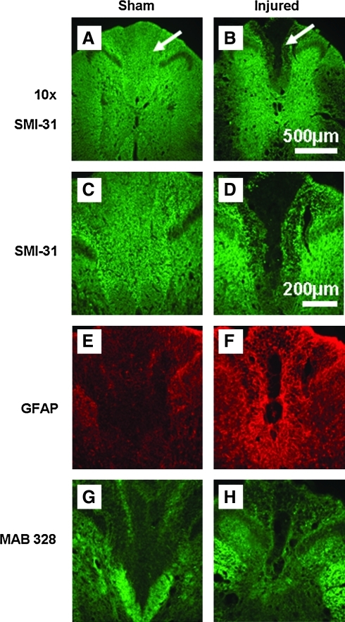
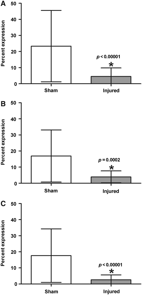
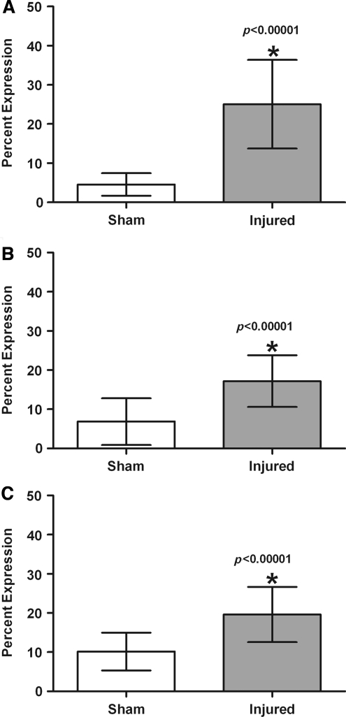
Comprehensive in vivo longitudinal studies that include multi-modal magnetic resonance imaging (MRI) and a battery of behavioral assays to assess functional outcome were performed at multiple time points up to 56 days post-traumatic spinal cord injury (SCI) in rodents. The MRI studies included high-resolution structural imaging for lesion volumetry, and diffusion tensor imaging (DTI) for probing the white matter integrity. The behavioral assays included open-field locomotion, grid walking, inclined plane, computerized activity box performance, and von Frey filament tests. Additionally, end-point histology was assessed for correlation with both the MRI and behavioral data. The temporal patterns of the lesions were documented on structural MRI. DTI studies showed significant changes in white matter that is proximal to the injury epicenter and persisted to day 56. White matter in regions up to 1 cm away from the injury epicenter that appeared normal on conventional MRI also exhibited changes that were indicative of tissue damage, suggesting that DTI is a more sensitive measure of the evolving injury. Correlations between DTI and histology after SCI could not be firmly established, suggesting that injury causes complex pathological changes in multiple tissue components that affect the DTI measures. Histological evidence confirmed a significant decrease in myelin and oligodendrocyte presence 56 days post-SCI. Multiple assays to evaluate aspects of functional recovery correlated with histology and DTI measures, suggesting that damage to specific white matter tracts can be assessed and tracked longitudinally after SCI.


Figures

FIG. 1.
FIG. 1.
Example of injury pathology in an MRI RARE image of a spinal cord 28 days post-SCI. Hypointense lesions correspond to hemorrhage and necrosis, while hyperintense lesions correspond to edema and demyelination (MRI, magnetic resonance imaging; RARE, rapid acquisition by relaxation enhancement; SCI, spinal cord injury).
FIG. 2.
FIG. 2.
Temporal profile of hyperintense and hypointense lesions in injured animals. (A) There was slight hyperintense injury in sham animals, but there were no significant changes in volume seen over the course of the study. (B) There was slight hypointense injury seen in sham animals, but there were no significant changes in volume throughout the 8 weeks. (C) In the injured animals on days 7 and 14 they had significantly more hyperintense lesioning than the more chronic time points at days 28, 42, and 56. (D) There were no significant differences in hypointense lesion volume between any of the time points.
FIG. 3.
FIG. 3.
Diffusion tensor imaging measures within lateral white matter at day 56. (A) Fractional anisotropy (FA) values in injured spinal cord tissue decrease with proximity to the injury epicenter. FA values at all points, up to 1 cm rostral and 1 cm caudal to the epicenter, are significantly less in injured animals. (B) Longitudinal diffusivity (λL) in the lateral region was lower in injured animals, but only significantly lower in region C1. (C) Transverse diffusivity (λT) values in the lateral region increased with proximity to the epicenter, indicating damage to myelin. λT Values in all regions were significantly higher than the values in control animals.
FIG. 4.
FIG. 4.
Diffusion tensor imaging values from region C3, 1 cm from the epicenter. (A) Injured animals had significantly lower fractional anisotropy (FA) values at days 7 (p =0.0002), 14 (p = 0.0009), 28 (p < 0.00001), 42 (p < 0.00001), and 56 (p < 0.00001). (B) Longitudinal diffusivity (λL) in the lateral region was significantly lower in injured animals at days 7 (p = 0.0014) and 28 (p < 0.00001). (C) Injured animals had significantly higher transverse diffusivity (λT) values at days 28 (p = 0.0005), 42 (p = 0.0012), and 56 (p < 0.00001).
FIG. 5.
FIG. 5.
Representative images of sham and injured tissue approximately 5 mm rostral to the epicenter. (A and B) The images were first obtained at 10 × (the images show SMI-31-stained tissue, and the arrows indicate the dorsal columns; scale bar = 500 μm). (CH) The dorsal, lateral, and ventral regions were individually assessed at 20 × (scale bar = 200 μm). Tissue was stained for neurofilament (C and D), astrocytes (E and F), and for myelin and oligodendrocytes (G and H; GFAP, GFAP, glial fibrillary acidic protein).
FIG. 6.
FIG. 6.
Quantitative analysis of neurofilament staining. Staining with the SMI-31 antibody was significantly decreased in injured animals in the (A) dorsal, (B) lateral, and (C) ventral regions at 56 days post-injury.
FIG. 7.
FIG. 7.
Quantitative analysis of myelin and oligodendrocyte staining. MAB 328 staining was significantly decreased in the (A) dorsal, (B) lateral, and (C) ventral regions of injured spinal cord at 56 days post-injury.
FIG. 8.
FIG. 8.
Quantitative analysis of astrocyte staining. Glial fibrillary acidic protein was significantly upregulated in the (A) dorsal, (B) lateral, and (C) ventral regions of injured tissue at 56 days post-injury compared to sham animals.

Similar articles

Cited by

References

    1. Basso D.M. Beattie M.S. Bresnahan J.C. A sensitive and reliable locomotor rating scale for open field testing in rats. J. Neurotrauma. 1995;12:1–21. - PubMed
    1. Basso D.M. Beattie M.S. Bresnahan J.C. Graded histological and locomotor outcomes after spinal cord contusion using the NYU weight-drop device versus transection. Exp. Neurol. 1996;139:244–256. - PubMed
    1. Behrmann D.L. Bresnahan J.C. Beattie M.S. Shah B.R. Spinal cord injury produced by consistent mechanical displacement of the cord in rats: behavioral and histologic analysis. J. Neurotrauma. 1992;9:197–217. - PubMed
    1. Bilgen M. Al-Hafez B. Alrefae T. He Y.Y. Smirnova I.V. Aldur M.M. Festoff B.W. Longitudinal magnetic resonance imaging of spinal cord injury in mouse: changes in signal patterns associated with the inflammatory response. Magn. Reson. Imaging. 2007;25:657–664. - PubMed
    1. Bockhorst K.H. Hui C.K. Narayana P.A. Fully automatic postprocessing and evaluation of DTI data: Unsupervised pipeline for batch jobs. 18th Annual Meeting of the International Society for Magnetic Resonance in Medicine; Stockholm, Sweden. 2010a.

Publication types

MeSH terms

Substances