. 2010 Aug;160(7):1699-713.
doi: 10.1111/j.1476-5381.2010.00828.x.

A novel, orally active LPA(1) receptor antagonist inhibits lung fibrosis in the mouse bleomycin model

Affiliations

A novel, orally active LPA(1) receptor antagonist inhibits lung fibrosis in the mouse bleomycin model

J S Swaney et al. Br J Pharmacol. 2010 Aug.

Abstract

Background and purpose: The aim of this study was to assess the potential of an antagonist selective for the lysophosphatidic acid receptor, LPA(1), in treating lung fibrosis We evaluated the in vitro and in vivo pharmacological properties of the high affinity, selective, oral LPA(1)-antagonist (4'-{4-[(R)-1-(2-chloro-phenyl)-ethoxycarbonylamino]-3-methyl-isoxazol-5-yl}-biphenyl-4-yl)-acetic acid (AM966).

Experimental approach: The potency and selectivity of AM966 for LPA(1) receptors was determined in vitro by calcium flux and cell chemotaxis assays using recombinant and native cell cultures. The in vivo efficacy of AM966 to reduce tissue injury, vascular leakage, inflammation and fibrosis was assessed at several time points in the mouse bleomycin model.

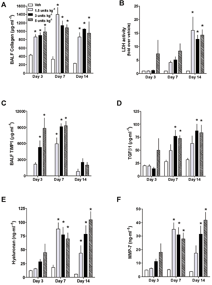
Key results: AM966 was a potent antagonist of LPA(1) receptors, with selectivity for this receptor over the other LPA receptors. In vitro, AM966 inhibited LPA-stimulated intracellular calcium release (IC(50)= 17 nM) from Chinese hamster ovary cells stably expressing human LPA(1) receptors and inhibited LPA-induced chemotaxis (IC(50)= 181 nM) of human IMR-90 lung fibroblasts expressing LPA(1) receptors. AM966 demonstrated a good pharmacokinetic profile following oral dosing in mice. In the mouse, AM966 reduced lung injury, vascular leakage, inflammation and fibrosis at multiple time points following intratracheal bleomycin instillation. AM966 also decreased lactate dehydrogenase activity and tissue inhibitor of metalloproteinase-1, transforming growth factor beta1, hyaluronan and matrix metalloproteinase-7, in bronchoalveolar lavage fluid.

Conclusions and implications: These findings demonstrate that AM966 is a potent, selective, orally bioavailable LPA(1) receptor antagonist that may be beneficial in treating lung injury and fibrosis, as well as other diseases that are characterized by pathological inflammation, oedema and fibrosis.


Figures

Figure 1
Figure 1
Pharmacological characterization of AM966 as an LPA1 receptor antagonist. (A) Structure of AM966. (B) Inhibition of LPA-stimulated intracellular calcium release from CHO cells stably expressing the mouse (mLPA1) and human LPA1 (hLPA1) receptors. CHO cells were pre-treated with increasing concentrations of AM966 for 30 min and then stimulated with LPA (10–30 nM) and calcium release was measured. (C) AM966-mediated inhibition of cell chemotaxis of human A2058 melanoma cells (IC50 = 138 nM), IMR-90 human lung fibroblasts (IC50 = 181 nM) and CHO mLPA1 cells (IC50 = 469 nM). LPA, lysophosphatidic acid; CHO, Chinese hamster ovary.
Figure 2
Figure 2
Time course of inflammatory, fibrotic and cell death markers in the mouse bleomycin model. Effects of bleomycin on (A) soluble collagen (B) LDH activity (cell death marker) and (C) TIMP-1 (D) TGFβ1 (E) hyaluronan and (F) MMP-7 concentrations in BALF at several time points (3, 7 and 14 days) following intratracheal instillation of bleomycin (1.5, 3, and 5 units·kg−1). Data represent the mean ± standard error of the mean of n≥ 4 mice per group. * denotes a significant increase (P < 0.05) compared to vehicle at each time point. LDH, lactate dehydrogenase; TGFβ-1, transforming growth factor β-1; TIMP-1, tissue inhibitor of metalloproteinase-1; MMP-7, matrix metalloproteinase-7; BALF, bronchoalveolar lavage fluid; Veh, vehicle.
Figure 3
Figure 3
Time-concentration profile for AM966. Fasted mice (n = 2) were dosed with AM966 and blood was sampled at 1, 2, 4, 8 and 24 h after oral dosing (10 mg·kg−1). The dashed line in Figure 3 represents the IC50 value for AM966-mediated inhibition of cellular chemotaxis by Chinese hamster ovary cells expressing recombinant mouse LPA1 receptors (See Figure 1D).
Figure 4
Figure 4
AM966 reduces vascular leakage, inflammation and lung injury and inflammation in a 3 day bleomycin model. Mice were given intratracheal bleomycin sulfate (BLM; 1.5 units·kg−1) or saline vehicle (Veh.), followed by oral (gavage) administration of AM966 (10 and 30 mg·kg−1, BID) for a period of 3 days. BALF was isolated and analysed for changes in (A) total protein, (B) inflammatory cells, and (C) LDH activity. Data represent the mean ± standard error of the mean of n = 5–7 mice per group. # denotes a significant increase (P < 0.05) compared to vehicle control. * denotes a significant decrease (P < 0.05) compared to BLM. BID, twice a day; BALF, bronchoalveolar lavage fluid; LDH, lactate dehydrogenase.
Figure 5
Figure 5
AM966 reduces vascular leakage and fibrosis in a 7 day bleomycin model. Mice were given intratracheal bleomycin sulfate (BLM; 3.0 units·kg−1) or saline vehicle (Veh.), followed by oral (gavage) administration of AM966 (1, 10 and 30 mg·kg−1, BID) for a period of 7 days. BALF was isolated and analysed for changes in (A) total protein, (B) collagen, (C) total (TGFβ1). (D) Representative histopathological images (100× magnification; trichrome staining) are shown of the lungs of mice treated with (a) vehicle, (b) BLM or (c) BLM + AM966 (30 mg·kg−1, BID). Blue staining denotes regions of fibrosis. (E) Total cellularity was analysed using a Hemavet system and differential cell counts for macrophages, neutrophils and lymphocytes were measured by cytospin in BALF. Data represent the mean ± standard error of the mean of n = 8 mice per group. # denotes a significant increase (P < 0.05) compared to vehicle control. * denotes a significant decrease (P < 0.05) compared to BLM. BID, twice a day; BALF, bronchoalveolar lavage fluid.
Figure 6
Figure 6
AM966 inhibits lung fibrosis and maintains body weight 14 days after bleomycin-induced lung injury. Mice were given intratracheal bleomycin sulfate (BLM; 1.5 units·kg−1) or saline vehicle (Veh.), followed by oral (gavage) administration of dexamethasone (1 mg·kg−1, QD) or AM966 (10, 30 and 60 mg·kg−1, BID) for a period of 14 days. (A, a-f) Representative histopathological images (100× magnification; Trichrome staining) are shown of the lungs of mice treated with (a) vehicle, (b) BLM, (c) BLM + dexamethasone (1 mg·kg−1, QD), (d) BLM + AM966 (10 mg·kg−1, BID), (e) BLM + AM966 (30 mg·kg−1, BID) or (f) BLM + AM966 (60 mg·kg−1, BID). (B) Soluble collagen in BALF. (C) Histopathological scoring of lung fibrosis. (D) Percentage change in mouse body weight (wt.). (E) Total cellularity and differential cell counts for macrophages, neutrophils and lymphocytes. Data represent the mean ± standard error of the mean of n = 8 mice per group. # denotes a significant increase (P < 0.05) compared to vehicle control. * denotes a significant decrease (P < 0.05) compared to BLM. QD, once a day; BID, twice a day; BALF, bronchoalveolar lavage fluid.
Figure 7
Figure 7
AM966 reduces vascular leakage, tissue injury and pro-fibrotic cytokine production. Mice were treated in the same manner as described for Figure 6. (A) Total protein (B) LDH activity (cell death marker) and concentrations of the pro-fibrotic factors (C) TIMP-1 (D) TGFβ1 (E) hyaluronan and (F) MMP-7 were analysed using commercially available kits. Data represent the mean ± standard error of the mean of n = 8 mice per group. # denotes a significant increase (P < 0.05) compared to vehicle control. Φ denotes a significant (P < 0.05) increase compared to BLM. * denotes a significant decrease (P < 0.05) compared to BLM. LDH, lactate dehydrogenase; TIMP-1, tissue inhibitor of metalloproteinase-1; TGFβ-1, transforming growth factor β-1; MMP-7, matrix metalloproteinase-7; BLM, bleomycin sulfate.
Figure 8
Figure 8
AM966 demonstrates greater efficacy than pirfenidone after i.t. instillation of bleomycin. Mice were given intratracheal bleomycin sulfate (BLM; 3.0 units·kg−1) or saline vehicle (Veh.) followed by oral administration of AM966 (30 mg·kg−1, BID) or pirfenidone (20, 100 and 400 mg·kg−1, BID) for a period of 14 days. Representative (A, panels a–f.) Trichrome-stained images (100× magnification) of lungs from mice treated with (a) vehicle, (b) BLM, (c) BLM + AM966 (30 mg·kg−1, QD), (d) BLM + pirfenidone (20 mg·kg−1, BID), (e) BLM + pirfenidone (100 mg·kg−1, BID) and (f) BLM + pirfenidone (400 mg·kg−1, BID). (B) soluble collagen concentrations in BALF (C) Histopathological scoring of lung fibrosis (D) Percentage change in mouse body weight (E) total protein in BALF (F) LDH activity and (G) TIMP-1 concentration are also shown. Data represent the mean ± standard error of the mean of n = 4–6 mice per group. # denotes a significant increase (P < 0.05) compared to vehicle control. * denotes a significant decrease (P < 0.05) compared to BLM. QD, once a day; BID, twice a day; BALF, bronchoalveolar lavage fluid; LDH, lactate dehydrogenase; TIMP-1, tissue inhibitor of metalloproteinase-1.
Figure 9
Figure 9
Effects of AM966 on mouse survival and lung fibrosis 21–28 days after BLM administration. (A) AM966 (30 mg·kg−1) significantly (P = 0.04) increased mouse survival over a 21 day period following i.t. instillation of BLM (5.0 units·kg−1). (B) AM966 (30 mg·kg−1) significantly (P < 0.05) decreased BALF collagen 28 days after i.t. instillation of BLM (3.0 units·kg−1). # denotes a significant increase (P < 0.05) compared to vehicle control. * denotes a significant decrease (P < 0.05) compared to BLM. BLM, bleomycin sulfate; BALF, bronchoalveolar lavage fluid.

References

    1. Alexander SPH, Mathie A, Peters JA. Guide to Receptors and Channels (GRAC), 4th edn. Br J Pharmacol. 2009;158(Suppl. 1):S1–S254. - PMC - PubMed
    1. Andersson-Sjoland A, de Alba CG, Nihlberg K, Becerril C, Ramirez R, Pardo A, et al. Fibrocytes are a potential source of lung fibroblasts in idiopathic pulmonary fibrosis. Int J Biochem Cell Biol. 2008;40:2129–2140. - PubMed
    1. Anliker B, Chun J. Cell surface receptors in lysophospholipid signaling. Semin Cell Dev Biol. 2004;15:457–465. - PubMed
    1. Ashcroft T, Simpson JM, Timbrell V. Simple method of estimating severity of pulmonary fibrosis on a numerical scale. J Clin Pathol. 1988;41:467–470. - PMC - PubMed
    1. Bjermer L, Lundgren R, Hallgren R. Hyaluronan and type III procollagen peptide concentrations in bronchoalveolar lavage fluid in idiopathic pulmonary fibrosis. Thorax. 1989;44:126–131. - PMC - PubMed

MeSH terms