Comparative Study
. 2010 Aug;160(8):1916-28.
doi: 10.1111/j.1476-5381.2010.00700.x.

Biochemical and molecular analysis of carboxylesterase-mediated hydrolysis of cocaine and heroin

Affiliations
Comparative Study

Biochemical and molecular analysis of carboxylesterase-mediated hydrolysis of cocaine and heroin

M J Hatfield et al. Br J Pharmacol. 2010 Aug.

Abstract

Background and purpose: Carboxylesterases (CEs) metabolize a wide range of xenobiotic substrates including heroin, cocaine, meperidine and the anticancer agent CPT-11. In this study, we have purified to homogeneity human liver and intestinal CEs and compared their ability with hydrolyse heroin, cocaine and CPT-11.

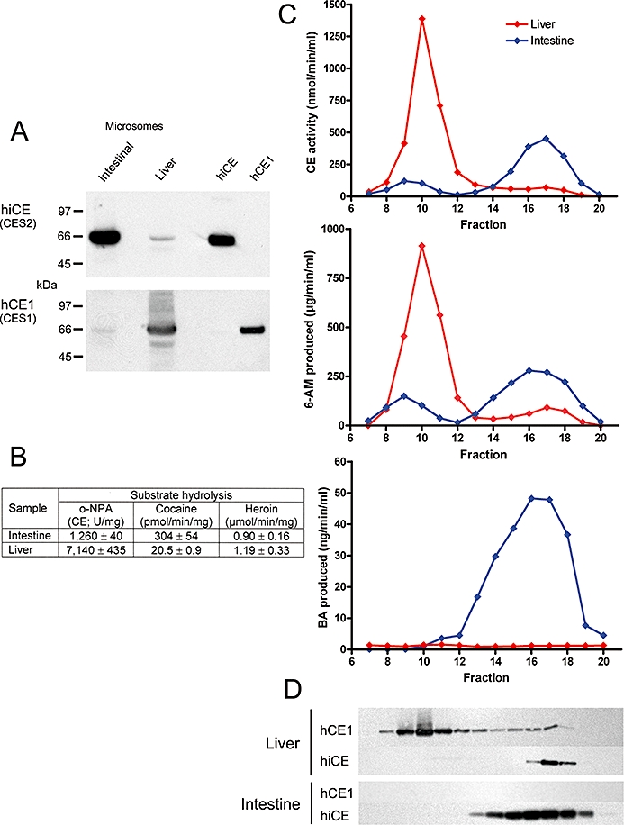
Experimental approach: The hydrolysis of heroin and cocaine by recombinant human CEs was evaluated and the kinetic parameters determined. In addition, microsomal samples prepared from these tissues were subjected to chromatographic separation, and substrate hydrolysis and amounts of different CEs were determined.

Key results: In contrast to previous reports, cocaine was not hydrolysed by the human liver CE, hCE1 (CES1), either as highly active recombinant protein or as CEs isolated from human liver or intestinal extracts. These results correlated well with computer-assisted molecular modelling studies that suggested that hydrolysis of cocaine by hCE1 (CES1), would be unlikely to occur. However, cocaine, heroin and CPT-11 were all substrates for the intestinal CE, hiCE (CES2), as determined using both the recombinant protein and the tissue fractions. Again, these data were in agreement with the modelling results.

Conclusions and implications: These results indicate that the human liver CE is unlikely to play a role in the metabolism of cocaine and that hydrolysis of this substrate by this class of enzymes is via the human intestinal protein hiCE (CES2). In addition, because no enzyme inhibition is observed at high cocaine concentrations, potentially this route of hydrolysis is important in individuals who overdose on this agent.


Figures

Figure 1
Figure 1
Sodium dodecyl sulphate-polyacrylamide gel electrophoresis analysis of different fractions following purification of hiCE (CES2). Approximately 3 µg of protein was loaded into each lane and following electrophoresis, the gel was stained with Coomassie Blue. Lane 1, Crude culture media; Lane 2, After ultrafiltration; Lane 3, After ion-exchange chromatography using DEAE; Lane 4, After ultrafiltration; Lane 5, After chromatography on Sephacryl S200 HR.
Figure 2
Figure 2
A graph demonstrating the linear relationships between the clogP values of the para-substituted nitrophenyl esters, and their observed Km constants for hiCE (CES2) (formula image
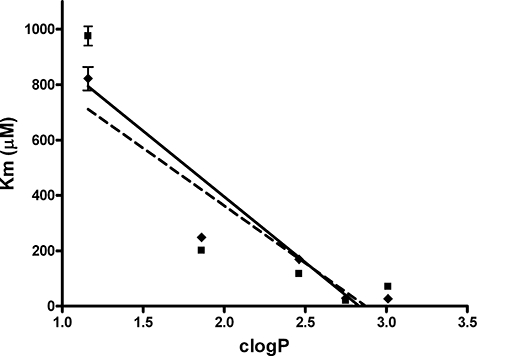
), and hCE1 (CES1) (formula image). The correlation coefficients (r2) for the data sets are indicated on the graph.
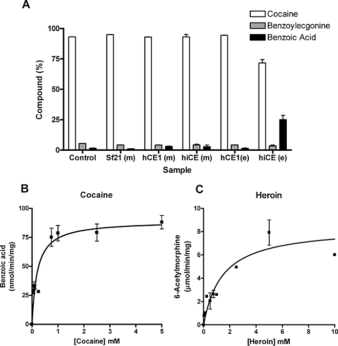
Figure 3
Figure 3
(A) Metabolism of cocaine by different carboxylesterases (CEs). Cocaine (1 mM) was incubated with the different samples for 1 h. The yields of benzoylecgonine and benzoic acid were determined by HPLC. Essentially, only hiCE (CES2) was able to hydrolyse cocaine. Control – 50 mM 4-(2-hydroxyethyl)-1-piperazineethanesulfonic acid (HEPES) buffer pH 7.4 only; Sf21 (m) – media harvested from uninfected Sf21 cells; hCE1 (m) – media harvested from Sf21 cells infected with baculovirus expressing hCE1 (CES1) (∼250 units of CE); hiCE (m) – media harvested from Sf21 cells infected with baculovirus expressing hiCE (CES2) (∼75 units of CE); hCE1 (e) – reaction containing pure hCE1 (CES1) (20 µg =∼2000 units of CE); hiCE (e) – reaction containing pure hiCE (CES2) (20 µg =∼500 units of CE). (B) The concentration versus velocity curve for hiCE (CES2) with cocaine. No evidence of enzyme inhibition by high concentrations of substrate was observed in these assays. (C) The concentration versus velocity curve for hiCE (CES2) with heroin. Similar to that seen for cocaine, no substrate-mediated enzyme inhibition was observed.
Figure 4
Figure 4
Chromatographic separation of carboxylesterases (CEs) in human intestinal and liver microsomal extracts using Sephacryl S-200 HR resin. (A) Western analysis demonstrating the amounts of hiCE (CES2) and hCE1 (CES1) present in the samples prior to chromatography. Standards present on the right hand side of the image represent 50 ng of each pure protein. (B) Table demonstrating the levels of CE activity, as well as the cocaine and heroin hydrolysis of the intestine and liver microsomal extracts prior to chromatography. (C) Elution profile for intestinal (blue line) and liver (red line) microsomal extracts demonstrating levels of CE activity (top graph), 6-acetylmorphine (6-AM) formation (heroin hydrolysis; middle graph) and benzoic acid (BA) production (cocaine hydrolysis; bottom graph). (D) Western analysis of these same fractions analysed in panel C, using antibodies specific for hiCE (CES2) or hCE1 (CES1). The images are aligned such that the signals correspond to the fraction number indicated on the abscissa axis.
Figure 5
Figure 5
Computer-assisted docking of cocaine or heroin into the active sites of hCE1 (CES1) or hiCE (CES2). (A) An overlay of the ribbon representations of the hCE1 (CES1) (taupe) and hiCE (CES2) (orange/brown) structures used for the docking studies. As can be seen one loop of hCE1 (CES1) (highlighted in pink) is displaced (marked by the white arrow) and projects towards the active site catalytic amino acids (Ser, His, Glu). (B) An overlay of the catalytic amino acids in hCE1 (CES1) (taupe) and hiCE (CES2) (orange/brown) with the distances between the relevant atoms indicated. (C, D) – Docking of cocaine into the active sites of hiCE (CES2) and hCE1 (CES1), respectively. The active site serine is depicted and the distance from the Oγ atom to the carbonyl carbon atoms in the drug are displayed. (E, F) – Docking of heroin into the active sites of hiCE (CES2) and hCE1 (CES1), respectively. The active site serine is depicted and the distance from the Oγ atom to the carbonyl carbon atoms in the drug are displayed. In all panels, the nitrogen and oxygen atoms displayed in blue and red, respectively, and distances are marked in Å.

References

    1. Anderson RN. Deaths: leading causes for 1999. Natl Vital Stat Rep. 1999;49:1–88. - PubMed
    1. Beaufay H, Amar-Costesec A, Feytmans E, Thines-Sempoux D, Wibo M, Robbi M, et al. Analytical study of microsomes and isolated subcellular membranes from rat liver. I. Biochemical methods. J Cell Biol. 1974;61:188–200. - PMC - PubMed
    1. Bencharit S, Morton CL, Hyatt JL, Kuhn P, Danks MK, Potter PM, et al. Crystal structure of human carboxylesterase 1 complexed with the Alzheimer's drug tacrine. From binding promiscuity to selective inhibition. Chem & Biol. 2003a;10:341–349. - PubMed
    1. Bencharit S, Morton CL, Xue Y, Potter PM, Redinbo MR. Structural basis of heroin and cocaine metabolism by a promiscuous human drug-processing enzyme. Nat Struct Biol. 2003b;10:349–356. - PubMed
    1. Bencharit S, Edwards CC, Morton CL, Howard-Williams EL, Kuhn P, Potter PM, et al. Multisite promiscuity in the processing of endogenous substrates by human carboxylesterase 1. J Mol Biol. 2006;363:201–214. - PMC - PubMed

Publication types

MeSH terms