Comparative Study
. 2010 Aug;160(8):1997-2007.
doi: 10.1111/j.1476-5381.2010.00814.x.

Nucleotide oligomerization domain 1 is a dominant pathway for NOS2 induction in vascular smooth muscle cells: comparison with Toll-like receptor 4 responses in macrophages

Affiliations
Comparative Study

Nucleotide oligomerization domain 1 is a dominant pathway for NOS2 induction in vascular smooth muscle cells: comparison with Toll-like receptor 4 responses in macrophages

L Moreno et al. Br J Pharmacol. 2010 Aug.

Abstract

Background and purpose: Gram-negative bacteria contain ligands for Toll-like receptor (TLR) 4 and nucleotide oligomerization domain (NOD) 1 receptors. Lipopolysaccharide (LPS) activates TLR4, while peptidoglycan products activate NOD1. Activation of NOD1 by the specific agonist FK565 results in a profound vascular dysfunction and experimental shock in vivo.

Experimental approach: Here, we have analysed a number of pharmacological inhibitors to characterize the role of key signalling pathways in the induction of NOS2 following TLR4 or NOD1 activation.

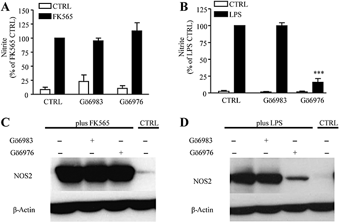
Key results: Vascular smooth muscle (VSM) cells expressed NOD1 mRNA and protein, and, after challenge with Escherichia coli or FK565, NOS2 protein and activity were induced. Macrophages had negligible levels of NOD1 and were unaffected by FK565, but responded to E. coli and LPS by releasing increased NO and expression of NOS2 protein. Classic pharmacological inhibitors for NF-kappaB (SC-514) and mitogen-activated protein kinase (SB203580, PD98059) signalling pathways inhibited responses in both cell types regardless of agonist. While TLR4-mediated responses in macrophages were specifically inhibited by the pan-caspase inhibitor z-VAD-fmk and the PKC inhibitor Gö6976, NOD1-mediated responses in VSM cells were inhibited by the Rip2 inhibitor PP2.

Conclusions and implications: Our findings suggest a selective role for NOD1 in VSM cells, and highlight NOD1 as a potential novel therapeutic target for the treatment of vascular inflammation.


Figures

Figure 1
Figure 1
Effects of Gram-negative PAMPs on NO release by cultured VSM cells and macrophages, and expression of NOD1 in VSM cells and macrophages. (A) At 24 h, Escherichia coli (EC; 3 × 10⁷ CFU·mL−1), LPS (1 µg·mL−1) or FK565 (10 nM) induced significant increases in release of NO from VSM cells. NO was measured by the oxidation product nitrite by Griess assay, and compared to cells incubated in media alone (CTRL). (B) Escherichia coli and LPS induced significant release of NO in J774.2 macrophages. In contrast, FK565 did not significantly increase the release of NO. Data are presented as the mean ± SEM from n= 6 determinations for J774.2 cells and n= 18 for VSM cells. (C,D) Representative Western blots of NOS2 (≈130 kDa) and β-actin (≈47 kDa) from VSM cells (C) and macrophages (D) treated with E. coli, LPS or FK565 for 24 h. (E) Representative Western blot of NOD1 protein (≈95 kDa) in VSM, J774.2 or freshly isolated peritoneal macrophages from C57 BL/6 mice. Detectable NOD1 expression was only seen in VSM cells. (F) Densitometric data analysis of NOD1 protein normalized to β-actin levels and expressed as arbitrary units (AU). Data are presented as the mean ± SEM from n= 3. (G) Ethidium bromide-stained agarose gels containing RT–PCR products of NOD1 (302 bp) and β-actin (467 bp) in VSM cells and J774.2 macrophages. Detectable NOD1 expression was only seen in VSM cells. (H) Densitometric data analysis of NOD1 mRNA normalized to β-actin levels and expressed as AU. Data are presented as the mean ± SEM from n= 3.
Figure 5
Figure 5
Effects of Src and serine/threonine kinase inhibitor PP2 on the induction of NOS2 by TLR4 and NOD1 agonists. Effects of the Src and serine/threonine kinase inhibitor PP2 (1 nM–1 µM) on the ability of FK565 or LPS to induce nitrite production in VSM cells (A) and macrophages (B). The data are the mean ± SEM of n= 9 for J774.2 cells and n= 15 for VSM cells, and are expressed as a percentage of the response to the FK565 or LPS alone (CTRL). (C) Representative image showing that 0.1 µM PP2 inhibited NOS2 induction in VSM cells by NOD1 activation (n= 3). Data were analysed using one-sample t-test for normalized data, *P < 0.05, **P < 0.01 and ***P < 0.001.
Figure 4
Figure 4
Role of protein kinase C isoforms (PKC) in TLR4 and NOD1 signalling pathways. VSM cells (A) and J774.2 macrophages (B) were treated with the PKC inhibitors Gö6983 (0.1 µM) and Gö6976 (0.1 µM). NOS2 activity was induced by FK565 (10 nM) in VSM cells and by LPS (1 µg·mL−1) in macrophages. Data are the mean ± SEM of n= 6 for J774.2 cells and n= 15 for VSM cells and are expressed as a percentage of the response to the FK565 or LPS alone (CTRL). Western blotting images for NOS2 protein (C,D) showing the effects of Gö6976 and Gö69683 (both at 0.1 µM) on NOS2 expression in VSM cells (C) and macrophages (D) (n= 3). Data were analysed using one-sample t-test for normalized data, *P < 0.05 and ***P < 0.001.
Figure 3
Figure 3
Role of MAPKs in TLR4 and NOD1 signalling pathways. Regulation of nitrite production in VSM cells (A, C, E, G) and macrophages (B, D, F, H) by the p38 MAPK inhibitor SB 203580 (0.01–10 µM), the ERK 1/2 inhibitor PD 98059 (0.01–100 µM) and the JNK inhibitors SP 600125 (0.1–30 µM) and D-JNKi (0.1–30 µM). Data are presented as the mean ± SEM of n= 6–9 for J774.2 cells, and n= 9–15 for VSM cells, and are expressed as a percentage of the response to the FK565 or LPS alone (CTRL). Representative Western blotting images for NOS2 protein (G,H) showing the effects of SB 203580 (1 µM), PD 98059 (10 µM) and SP 600125 (30 µM) on NOS2 expression in VSM cells (G) and macrophages (H). Data were analysed using one-sample t-test for normalized data, *P < 0.05 and ***P < 0.001, n= 3–4.
Figure 2
Figure 2
Role of corticosteroids and NF-κB on TLR4 and NOD1-induced NO production and NOS2 expression in VSM and J774 cells. VSM cells (A,E) and J774.2 macrophages (B,F) were treated with increasing concentrations of dexamethasone (0.01–10 µM; A,B) or the IKK 2 inhibitor SC-514 (0.1–30 µM; E,F). NOS2 was induced in VSM cells with FK565 (10 nM) and in macrophages with LPS (1 µg·mL−1). Data were collected from n= 9–12 for J774.2 cells and n= 6 for VSM cells and expressed as a percentage of the response induced by FK565 or LPS, respectively (CTRL). Western blotting (C,D,G,H) for NOS2 protein showed that dexamethasone (1 µM; C,D) and SC-514 (10 µM; G,H) inhibited protein expression in both VSM cells (C,E) and macrophages (D,H) at 24 h (n= 3–4). Data were analysed using one-sample t-test for normalized data, *P < 0.05, **P < 0.01 and ***P < 0.001.
Figure 6
Figure 6
Role of the inflammasome in the induction of NOS2 by TLR4 and NOD1 agonists. (A,B) Effects of the pan-caspase inhibitor Z-VAD-fmk (0.1–30 µM) on the ability of FK565 or LPS to induce nitrite production in VSM cells (A) and macrophages (B). (C,D) Representative Western blotting images showing the effects of Z-VAD-fmk (10 µM) on NOS2 induction in VSM cells (C) and J774.2 macrophages (D) (n= 3–6). (E) Increasing concentrations of anti-IL1β- and anti-IL18-neutralizing antibodies (10−8 to 10−5 g·mL−1) did not inhibit NOS2 activity in VSM cells stimulated with FK565. (F) The selective caspase-1 inhibitor Z-WEDH-fmk and the caspase-8 inhibitor Z-IETD-fmk (both at 0.1–30 µM) similarly inhibited NOS2 induction by the TLR4 agonist LPS in macrophages. The data are the mean ± SEM of n= 6–9 for VSM cells and n= 6–12 for J774.2 cells, and are expressed as a percentage of the response to the FK565 or LPS alone (CTRL). Data were analysed using one-sample t-test for normalized data, *P < 0.05, **P < 0.01 and ***P < 0.001.

References

    1. Argast GM, Fausto N, Campbell JS. Inhibition of RIP2/RIck/CARDIAK activity by pyridinyl imidazole inhibitors of p38 MAPK. Mol Cell Biochem. 2005;268:129–140. - PubMed
    1. Asehnoune K, Strassheim D, Mitra S, Yeol Kim J, Abraham E. Involvement of PKCalpha/beta in TLR4 and TLR2 dependent activation of NF-kappaB. Cell Signal. 2005;17:385–394. - PubMed
    1. Bennett BL, Sasaki DT, Murray BW, O'Leary EC, Sakata ST, Xu W, et al. SP600125, an anthrapyrazolone inhibitor of Jun N-terminal kinase. Proc Natl Acad Sci U S A. 2001;98:13681–13686. - PMC - PubMed
    1. Bishop-Bailey D, Larkin SW, Warner TD, Chen G, Mitchell JA. Characterization of the induction of nitric oxide synthase and cyclo-oxygenase in rat aorta in organ culture. Br J Pharmacol. 1997;121:125–133. - PMC - PubMed
    1. Borsello T, Bonny C. Use of cell-permeable peptides to prevent neuronal degeneration. Trends Mol Med. 2004;10:239–244. - PubMed

Publication types

MeSH terms

Substances