. 2010 Aug;160(8):2008-27.
doi: 10.1111/j.1476-5381.2010.00861.x.

Combretastatin A-4 inhibits cell growth and metastasis in bladder cancer cells and retards tumour growth in a murine orthotopic bladder tumour model

Affiliations

Combretastatin A-4 inhibits cell growth and metastasis in bladder cancer cells and retards tumour growth in a murine orthotopic bladder tumour model

Cheng-Huang Shen et al. Br J Pharmacol. 2010 Aug.

Abstract

Background and purpose: Bladder cancer is a highly recurrent cancer after intravesical therapy, so new drugs are needed to treat this cancer. Hence, we investigated the anti-cancer activity of combretastatin A-4 (CA-4), an anti-tubulin agent, in human bladder cancer cells and in a murine orthotopic bladder tumour model.

Experimental approach: Cytotoxicity of CA-4 was measured by 3-(4,5-dimethylthiazol-2-yl)-2,5-diphenyltetrazolium bromide (MTT) assay, propidium iodide (PI) staining assay and clonogenic survival assay. In vivo microtubule assembly assay, cell cycle analyses, Western blot and cell migration assay were used to study the mechanism of CA-4. The effect of intravesical CA-4 therapy on the development of tumours was studied in the murine orthotopic bladder tumour model.

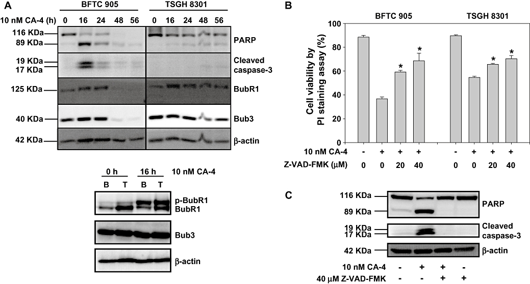
Key results: CA-4 inhibited microtubule polymerization in vivo. Cytotoxic IC(50) values of CA-4 in human bladder cancer cells were below 4 nM. Analyses of cell-cycle distribution showed CA-4 obviously induced G(2)-M phase arrest with sub-G(1) formation. The analyses of apoptosis showed that CA-4 induced caspase-3 activation and decreased BubR1 and Bub3 in cancer cells. In addition to apoptosis, CA-4 was also found to induce the formation of multinucleated cells. CA-4 had a significantly reduced cell migration in vitro. Importantly, the in vivo study revealed that intravesical CA-4 therapy retarded the development of murine bladder tumours.

Conclusions and implications: These data demonstrate that CA-4 kills bladder cancer cells by inducing apoptosis and mitotic catastrophe. It inhibited cell migration in vitro and tumour growth in vivo. Hence, CA-4 intravesical therapy could provide another strategy for treating superficial bladder cancers.


Figures

Figure 1
Figure 1
Structure of CA-4 and its inhibitory effect on microtubule polymerization in vivo. (A) Chemical structure and HPLC analyses data of synthetic cis CA-4. (B and C) Inhibitory effect of CA-4 on microtubule polymerization in vivo. BFTC 905 (B) and TSGH 8301 (C) cells were treated with CA-4 or with the same volume of DMSO as control. After 6 h incubation, cells were lysed in lysis buffer. Cell lysates were centrifuged to separate polymerized microtubules from free tubulin dimers as described in Methods. β-Actin was used as a loading control. The columns represent the means from three independent experiments and presented as mean ± SD. *P < 0.05, a significant difference compared with the control cells. CA-4, combretastatin A-4; DMSO, dimethylsulphoxide.
Figure 2
Figure 2
(A) Cytotoxicity of CA-4 against two human bladder cancer cell lines and a normal human uroepithelium. BFTC 905 and TSGH 8301 cells were initially seeded at 9x10³ cells per well and SV-HUC-1 was seeded at 1.5x10⁴ cells per well in 96-well dishes and then treated with various concentrations of CA-4 or DMSO for 48 h. The cell viability was measured by MTT assay. Measurement was performed in quadruplicate and presented as mean ± SD. This experiment was repeated three times. (B) CA-4-induced cell death in two human bladder cancer cell lines and a normal human uroepithelium. BFTC 905 and TSGH 8301 cells were initially seeded at 8x10⁵ cells and SV-HUC-1 was seeded at 2.8x10⁶ cells in 100-mm dishes and then treated with various concentrations of CA-4 or DMSO for 24 h and 48 h. Then the cells were collected and stained with PI for flow cytometry analyses. The columns represent the means from three independent experiments and are presented as mean ± SD. (C) Effect of CA-4 on clonogenic survival assay. BFTC 905 and TSGH 8301 cells were initially seeded at 1x10³ cells in 100-mm dishes and then treated with various concentrations of CA-4 or DMSO for 1 h. After being rinsed with fresh medium, cells were allowed to grow for 14 days to form colonies. The columns represent the means of triplicate experiment and are presented as mean ± SD. *P < 0.05, a significant difference compared with the control cells. CA-4, combretastatin A-4; DMSO, dimethylsulphoxide; MTT, 3-(4,5-dimethylthiazol-2-yl)-2,5-diphenyltetrazolium bromide; PI, propidium iodide.
Figure 3
Figure 3
(A) Effect of CA-4 on cell cycle distribution in bladder cancer cells. BFTC 905 and TSGH 8301 cells were treated with 10 nM CA-4 or DMSO for 24 h or 48 h, then the cells were collected for flow cytometry cell cycle analyses. (B) The expression of MPM-2 after CA-4 treatment. BFTC 905 and TSGH 8301 cells were treated with various concentrations of CA-4 or DMSO for 24 h, then the cells were collected and lysed for Western blot. The columns represent the means from three independent experiments and are presented as mean ± SD. *P < 0.05, a significant difference compared with the control cells. (C) Effect of CA-4 on cell cycle distribution in SV-HUC-1 cells. Cells were treated with 10 nM CA-4 or DMSO for 24 h or 48 h, then the cells were collected for flow cytometry cell cycle analyses. CA-4, combretastatin A-4; DMSO, dimethylsulphoxide.
Figure 3
Figure 3
(A) Effect of CA-4 on cell cycle distribution in bladder cancer cells. BFTC 905 and TSGH 8301 cells were treated with 10 nM CA-4 or DMSO for 24 h or 48 h, then the cells were collected for flow cytometry cell cycle analyses. (B) The expression of MPM-2 after CA-4 treatment. BFTC 905 and TSGH 8301 cells were treated with various concentrations of CA-4 or DMSO for 24 h, then the cells were collected and lysed for Western blot. The columns represent the means from three independent experiments and are presented as mean ± SD. *P < 0.05, a significant difference compared with the control cells. (C) Effect of CA-4 on cell cycle distribution in SV-HUC-1 cells. Cells were treated with 10 nM CA-4 or DMSO for 24 h or 48 h, then the cells were collected for flow cytometry cell cycle analyses. CA-4, combretastatin A-4; DMSO, dimethylsulphoxide.
Figure 4
Figure 4
(A) Effect of CA-4 on the expression of apoptotic proteins and M phase checkpoint proteins. BFTC 905 (B) and TSGH 8301 (T) cells were treated with 10 nM CA-4 or DMSO for indicated times, then the cells were collected and lysed for Western blot. (B) Effect of Z-VAD-FMK on CA-4-induced cell death. BFTC 905 and TSGH 8301 cells were pretreated with or without different concentrations of Z-VAD-FMK for 1 h before being exposed to 10 nM CA-4 or DMSO for 24 h. The cell viability was measured by PI staining assay. The columns represent the means from three independent experiments and presented as mean ± SD. *P < 0.05, a significant difference compared with the CA-4-treated cells. (C) Effect of Z-VAD-FMK on CA-4-induced PARP and caspase 3 cleavage. BFTC 905 cells were pretreated with or without 40 µM Z-VAD-FMK for 1 h before being exposed to 10 nM CA-4 or DMSO for 24 h, then the cells were collected and lysed for Western blot. CA-4, combretastatin A-4; DMSO, dimethylsulphoxide.
Figure 5
Figure 5
(A) CA-4 induced giant multinucleation in BFTC 905 and TSGH 8301 cells. Cells were treated with DMSO or 10 nM CA-4 for 24 h, and then stained with H33342 to label DNA. The light (left) and fluorescent (right) images of BFTC 905 and TSGH 8301 cells are presented. (B) Calculation of CA-4-induced multinucleation in BFTC 905 and TSGH 8301 cells. Representative light (left) and fluorescent (right) images of BFTC 905 and TSGH 8301 cells after 10 nM CA-4 treatment for 24 h. Arrows indicate binucleated or multinucleated cells. The scale bar 100 um indicates 100 micrometers. Lower panel shows tha number of multinucleated cells from three independent experiments. *P < 0.05, a significant difference between the control and the CA-4-treated cells. CA-4, combretastatin A-4; DMSO, dimethylsulphoxide.
Figure 5
Figure 5
(A) CA-4 induced giant multinucleation in BFTC 905 and TSGH 8301 cells. Cells were treated with DMSO or 10 nM CA-4 for 24 h, and then stained with H33342 to label DNA. The light (left) and fluorescent (right) images of BFTC 905 and TSGH 8301 cells are presented. (B) Calculation of CA-4-induced multinucleation in BFTC 905 and TSGH 8301 cells. Representative light (left) and fluorescent (right) images of BFTC 905 and TSGH 8301 cells after 10 nM CA-4 treatment for 24 h. Arrows indicate binucleated or multinucleated cells. The scale bar 100 um indicates 100 micrometers. Lower panel shows tha number of multinucleated cells from three independent experiments. *P < 0.05, a significant difference between the control and the CA-4-treated cells. CA-4, combretastatin A-4; DMSO, dimethylsulphoxide.
Figure 6
Figure 6
(A) Effect of CA-4 on cell migration. BFTC 905 or TSGH 8301 cells were seeded on the upper chamber of the transwell system for 24 h. The upper chamber media were replaced by serum-free media with various concentrations of CA-4 or DMSO, and the lower chamber was filled with 10% FBS media. After a 24 h incubation period, the cells remaining on the upper surface of the filter membrane were removed, and the cells on the opposite surface of the filter membrane were stained with crystal violet and photographed under microscopy. The histograms show the number of migrated cells. (B) Effect of CA-4 on AKT phosphorylation. Human bladder cancer cell lines were untreated or treated with various concentrations of CA-4 for 24 h. Cells were harvested and the cell extract was prepared and loaded on SDS-PAGE. β-Actin was used as a loading control. The means from three independent experiments are presented as mean ± SD in the histograms. *P < 0.05, a significant difference versus the control cells. CA-4, combretastatin A-4; DMSO, dimethylsulphoxide.
Figure 6
Figure 6
(A) Effect of CA-4 on cell migration. BFTC 905 or TSGH 8301 cells were seeded on the upper chamber of the transwell system for 24 h. The upper chamber media were replaced by serum-free media with various concentrations of CA-4 or DMSO, and the lower chamber was filled with 10% FBS media. After a 24 h incubation period, the cells remaining on the upper surface of the filter membrane were removed, and the cells on the opposite surface of the filter membrane were stained with crystal violet and photographed under microscopy. The histograms show the number of migrated cells. (B) Effect of CA-4 on AKT phosphorylation. Human bladder cancer cell lines were untreated or treated with various concentrations of CA-4 for 24 h. Cells were harvested and the cell extract was prepared and loaded on SDS-PAGE. β-Actin was used as a loading control. The means from three independent experiments are presented as mean ± SD in the histograms. *P < 0.05, a significant difference versus the control cells. CA-4, combretastatin A-4; DMSO, dimethylsulphoxide.
Figure 7
Figure 7
Effect of CA-4 in a murine orthotopic bladder tumour model. (A) The time schedule for the murine orthotopic bladder tumour model. (B) The urinary system of mice with no tumour implantated and those treated with vehicle or CA-4 after tumour implantation. After the mice had been killed, the urinary system was isolated and photographed. Arrows indicate the bladders of mice with no tumour implanted (left), tumour-implanted mice treated with vehicle (middle) or treated with 50 mg·kg−1 CA-4 (right). ‘K’ indicates the kidney. (C) The bladder volume of a mouse with no tumour implanted and those with tumours treated with vehicle or CA-4. After the mice had been killed, the bladder volume was calculated by (length × width2)/2. *P < 0.05, a significant difference between the vehicle-treated and the CA-4-treated mice. (D) Bladder sections stained with haematoxylin and eosin (H&E) for histopathology. Magnification: 100x. (i) The bladder tissue of mice without a tumour after intravesical CA-4 therapy. ‘M’ stands for muscle layer. (ii) The bladder tissue of mice with a tumour and treated with 50 mg·kg−1 CA-4, the arrow indicates the edge of the tumour. (iii) The bladder tissue of mice with a tumour and treated with vehicle alone, the arrow indicates the outer edge of the bladder. (iv) The tumour in a vessel outside the bladder of the mice after tumour implantation and treatment with vehicle alone, the arrow indicates the tumour thrombosis. CA-4, combretastatin A-4.

Similar articles

Cited by

References

    1. Amling CL. Diagnosis and management of superficial bladder cancer. Curr Probl Cancer. 2001;25:219–278. - PubMed
    1. Barocas DA, Clark PE. Bladder cancer. Curr Opin Oncol. 2008;20:307–314. - PubMed
    1. Bilenker JH, Flaherty KT, Rosen M, Davis L, Gallagher M, Stevenson JP, et al. Phase I trial of combretastatin a-4 phosphate with carboplatin. Clin Cancer Res. 2005;11:1527–1533. - PubMed
    1. Black PC, Dinney CP. Bladder cancer angiogenesis and metastasis – translation from murine model to clinical trial. Cancer Metastasis Rev. 2007;26:623–634. - PubMed
    1. de Bruin EC, Medema JP. Apoptosis and non-apoptotic deaths in cancer development and treatment response. Cancer Treat Rev. 2008;34:737–749. - PubMed

Publication types

MeSH terms